









Testicular Cancer
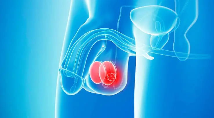
The testes are part of the male reproductive system and are located in the scrotum (testicles). Their primary function is to secrete the male hormone (testosterone) and produce reproductive cells (sperm). Testicular cancer is a tumor that begins in the testes and can sometimes spread to other areas of the body. It accounts for 1% of cancers in men, and its incidence is increasing.
What is testicular cancer?
The testes are part of the male reproductive system and are located in the scrotum (testicles). Their primary function is to secrete the male hormone (testosterone) and produce reproductive cells (sperm). Testicular cancer is a tumor that begins in the testes and can sometimes spread to other areas of the body. It accounts for 1% of cancers in men, and its incidence is increasing. It is more endemic in Northern European and Scandinavian countries. It is more frequently observed in children from families with higher socioeconomic and cultural levels. The right testicle is more often affected, as the risk factor, undescended testicle pathology, is more common on the right side. In rare cases, it can be bilateral (2-3%). The lifetime risk of developing a testicular tumor is 0.2%.
Types of Testicular Cancer (Pathology)
Testicular tumors have numerous histopathological types. The most common testicular tumors encountered clinically are germinal cell tumors. 90-95% of testicular tumors are germinal cell tumors. The remainder are non-germinal tumors. During embryonic development, if totipotential germ cells differentiate normally, spermatocytes are formed. If these totipotential germ cells differentiate abnormally, seminoma or embryonal carcinoma develops. If embryonal cells differentiate further following intraembryonic development, teratoma develops. If embryonal cells differentiate further following extraembryonic development, choriocarcinoma or yolk sac tumors form. Germinal cell testicular cancers are divided into two types, seminomatous and nonseminomatous cell testicular cancers, due to differences in their treatments.
Seminoma is a testicular tumor that can occur in men of all ages, but is more common in men aged 30-40 and generally has a better prognosis compared to other testicular tumors.
Non-seminoma testicular tumors include embryonal carcinoma, yolk sac tumor, choriocarcinoma, and teratoma. These can occur alone or, more often, in combination with seminoma, presenting as mixed tumors. They are more aggressive and prone to complications in the 18-35 age group.
- Seminom:Seminoma is the most common tumor among testicular cancers. While it can occur in men of all ages, it is more common in men aged 30-40 and generally has a better prognosis compared to other testicular tumors. Its incidence alone is 35%. There are three types of seminoma: classic seminoma, anaplastic seminoma, and spermatocytic seminoma. From a prognostic perspective, there is no difference between the three. 85% of seminomas are of the classic type. 10-15% contain syncytiotrophoblastic elements. In this type of seminoma, hCG levels are high in the blood due to hCG production. 5-10% of seminomas are anaplastic type. 5-10% of seminomas are spermatocytic type and are more common in men over 50 years of age.
- Embryonic Carcinoma:Its isolated occurrence rate is 20%. It is a more aggressive and progressive testicular cancer that occurs in the 18-35 age group. There are two types: adult and infantile. The infantile type is also called endodermal sinus tumor or yolk sac tumor.
- Teratoma:The probability of it occurring alone is 5%. There are two types: mature teratoma and immature teratoma. Mature teratomas contain three basic embryonic layers: ectoderm, mesoderm, and endoderm; therefore, these tumors contain structures that mimic all kinds of organs (bone, skin, teeth, and other organ cells).
- Koriyokarsinom:Its isolated occurrence rate is less than 1%. It is a highly aggressive tumor. It differs from other non-seminomatous testicular tumors due to its ability to metastasize early hematogenously.
- Mixed cell tumors:These tumors account for approximately 40% of cases and are the most common type of testicular cancer. About three-quarters of these mixed tumors are teratocarcinomas. Teratocarcinoma is a combination of teratoma and embryonal carcinoma. In approximately 6% of these tumors, seminoma is always present within the combination. The higher the percentage of embryonal carcinoma in mixed tumors, the worse the prognosis.
- Carcinoma in situ:In 5.2% of patients with testicular tumors, carcinoma in situ is found in the other testicle. These have a risk of later developing into invasive cancer.
Testicular Cancer Risk Factors, What Causes Testicular Cancer?
- Ages 18 to 35
- A history of undescended testicles (cryptorchidism) (a proven risk factor): 7-10% of patients with testicular tumors have a history of undescended testicles. Several factors are implicated in the development of cancer in undescended testicles. These include abnormal germ cell morphology, high temperature, impaired testicular blood supply, endocrine dysfunction (hormonal dysfunction), and gonadal dysgenesis (genital developmental disorder). The risk of developing testicular tumors in patients with undescended testicles is 3 to 14 times higher than expected. Approximately 5 to 10% of patients with undescended testicles have a risk of developing a tumor in the opposite testicle in its normal location.
- Hypospadias (also known as hypospadias) is a congenital genital developmental disorder in which the urethral opening is located in an area other than its normal position.
- Infertility is caused by significant abnormalities in sperm count and morphology.
- Abnormal testicular development, having atrophic (small) testicles.
- Family history, gene alterations on chromosome Xq27, and a history of testicular cancer (proven risk factors)
- Being of the White Race
- Genetic factors (the role of genes such as Bcl-2, p53, EGF, Rb)
- Testicular microlithiasis (calcifications)
- Excess endogenous or exogenous estrogen in the mother. It is known that when estrogen is given externally to pregnant mothers, the risk of testicular cancer in male babies increases significantly.
- Testicular traumas
- Sociocultural and economic level
Symptoms and Complaints
Abnormal growth in the testicles,palpable mass in the testicleThe most common finding and complaint is the feeling and detection of a hard mass/lump. However, not every mass or lump felt by hand is cancerous. In addition, a painful mass in the testicle, pain in the testicle and scrotum (10-15%) may be felt. If testicular cancer has spread to other parts of the body (metastasis), dull pain or some hardness may be felt in these areas. Hydrocele may be detected in 10-15% of patients along with the tumor. The patient can examine himself with his hands, especially after a warm bath, compare the two testicles with each other, and should definitely consult a doctor if he feels an abnormal size, hardness, or mass. Patients may present with complaints related to the areas where metastasis has occurred, such as cough, shortness of breath (lung metastasis), abdominal pain, loss of appetite, nausea and vomiting (retroperitoneal metastasis).
Diagnosis
After a thorough examination of the patient's history and medical background, ultrasonography and a series of blood tests, including important tumor markers, are required. Biopsy of testicular tumors is generally not recommended; it is usually avoided except in very special circumstances. In some cases, biopsy may be considered for frozen section pathological examination during surgery.
Blood Tests:Essentially, it includes tumor markers such as Beta-Human chorionic gonadotropin (Beta-HCG), Alpha-fetoprotein (AFP), and Lactic Dehydrogenase (LDH). Elevated levels of one or more of these are used to determine the type of cancer, make treatment decisions, and monitor patients. In non-seminoma tumors, 40-70% show elevated AFP or hCG levels, and 20-60% show elevated LDH levels. In seminomas, 15-20% show elevated hCG levels, and 20-60% show elevated LDH levels. AFP levels are not elevated in pure seminoma and choriocarcinoma. Additionally, markers such as Placental Alkaline Phosphatase (PLAP), Neuron-Specific Enolase (NSE), and Gamma-glutamyl transpeptidase (GGT) are sometimes used.
Ultrasonography (USG):Ultrasound provides invaluable information about the structure, nature, size, relationship with other tissues, and extent of a mass in the testicle. It can also detect microcalcifications (microlithiasis “microliths” – sandy and microcalcifications) which are considered precursors and risk factors for testicular cancer. Ultrasound is considered a fundamental imaging technique for detecting testicular cancer and can also be used to visualize metastases.
Magnetic resonance imaging (MRI):Detecting a testicular mass provides information about its structure and nature, and also guides us in detecting and staging metastases. It can also serve as a guide for biopsy sampling.
Spread of Testicular Cancer
Testicular cancers, except for choriocarcinoma, metastasize via the lymphatic route. Choriocarcinoma metastasizes hematogenously, to atypical locations, and at an early stage, and has a poorer prognosis compared to other types. Testicular tumors primarily metastasize to lymph nodes between T1 and L4, and around the major arteries surrounding the kidney (aorta and vena cava). The testis shares a congenital origin with the kidney, and metastases occur in these areas. Initially, metastases are located in this region, then gradually spread upwards to the mediastinum of the lungs and supraclavicular lymph nodes. While metastases are occurring upwards, lymphatic spread also occurs downwards, to the iliac lymph nodes. In advanced stages, metastases to the lungs, liver, bones, and brain are observed.
Stages of Testicular Cancer
Following high radical orchiectomy surgery, the histological structure and characteristics of the tumor are determined for disease staging. Postoperative tumor markers (HCG, AFP) values are re-evaluated. CT scans of the lungs, abdomen, and pelvis are performed. The Royal Marsden classification or the American Joint Committee on Cancer (AJCC) TNM classification is used. This classification shows the stages and level of tumor spread in the abdomen, guiding the treatment and follow-up of the cancer. Here, the classification is based on the extent of the primary tumor focus (T), the spread to the lymph nodes and the degree of spread (L), distant organ metastases (M), and the presence and level of tumor markers (S). After this information, testicular cancers are staged as follows:
Staging according to the Royal Marsden classification:
- Evre I:Cancer confined to the testicle; no tumor detected in other areas.
- Hebrews 2:Lymphatic spread is present (a: spread to intra-abdominal lymphatics smaller than 2 cm; b: spread to lymph nodes larger than 2-5 cm; c: lymphatic spread forming a mass larger than 5 cm).
- Evre III:Spread to the lymph nodes above the diaphragm
- Stage IV:Distant organ metastasis is present (lungs, liver, bones, brain, etc.) / spread via the bloodstream.
Differential Diagnosis of Testicular Cancer
Testicular tumors are easily confused with other intrascrotal diseases. Therefore, approximately 25% of patients with testicular tumors are misdiagnosed during their initial examination. Diseases that can be confused with testicular cancer include epididymitis or orchioepididymitis, hydrocele (which can sometimes be seen in conjunction with testicular tumors in 10-15%), spermatocele, hematocele, varicocele, tuberculous orchitis, and epidermoid cysts.
Prognosis/Course
Testicular cancer has a better prognosis and response to treatment compared to other organ cancers, and the chance of cure is higher, especially when caught in the early stages. As mentioned, the prognosis in testicular cancer depends on the stage of the disease, the cell type (seminoma or non-seminoma), the presence or absence of distant organ metastasis, the levels of serum markers, especially in non-seminoma cancers, and your individual and family history of testicular tumors. The higher the rate of embryonal carcinoma in non-seminomatous tumors, the worse the prognosis. Conversely, the presence of yolk sac tumors indicates a relatively good prognosis. Primary choriocarcinoma occurs at a younger age and has a relatively poor prognosis. Patients are divided into certain risk groups, and their treatment and follow-up are arranged accordingly. A patient with no poor prognostic factors is considered low-risk, a patient with 1 or 2 poor risk factors is considered moderate-risk, and a patient with 3-4 factors is considered high-risk.
Risk factors include: the histological presence of embryonal carcinoma in orchiectomy material is considered a poor prognostic risk factor, and even the proportion of embryonal cancer in the total tumor is significant. Furthermore, the presence of yolk sac tumor is considered a good prognostic factor, while the presence of lymphatic and/or vascular invasion is considered a poor risk factor.
Testicular Cancer Treatment
A multidisciplinary approach in the treatment of testicular cancer.
Decision-making in cancer treatment requires combining a large amount of data, including symptoms and imaging information such as MRI/CT scans and PET scans, and necessitates a multidisciplinary approach—integration of all relevant departments—to select the most appropriate treatment for the patient. International standards in cancer treatment require this approach.
At Prof. Dr. Hakkı Perk's private clinic, treatment planning for uro-oncological, or cancer, patients is carried out using a multidisciplinary approach. Once a week, at a hospital with which I have an agreement, cancer patients are discussed in an oncological council attended by urologists, medical oncologists, radiation oncology specialists, nuclear medicine specialists, pathologists, and radiologists. Treatment decisions and processes for these patients are finalized. Treatment processes for patients with prostate cancer, kidney cancer, adrenal gland tumors, bladder cancer, and testicular cancer are determined with the participation of all relevant departments.
Furthermore, cancer patients are guided by international oncology guidelines such as NCCN, ASCO, EAU, and AUA in their treatment management. In such multidisciplinary collaborations and cancer councils, all aspects of a patient's condition are considered and decided upon. Feedback presentations regarding patients undergoing treatment are also given at these councils.
Orchiectomy:The first-line treatment for testicular cancer is surgical removal of the cancerous testicle and its appendages (epididymis, spermatic cord, and funuculus). If there is any doubt in the diagnosis, frozen section pathology is performed during surgery if necessary, and a decision is made accordingly. In very special cases (bilateral tumor, patient having only one testicle, etc.), only the cancerous part of the testicle may be removed. Testicular surgery is performed through an incision higher up in the groin, not in the scrotum (high inguinal orchiectomy). The tissue is sent to pathology, and the need for additional treatment is determined based on the results of pathology and imaging studies. Additional treatments include further surgical procedures, chemotherapy, and radiotherapy. Testicular cancers respond quite well to radiotherapy and chemotherapy. After testicular removal, a testicular prosthesis can be implanted in the same procedure or later for cosmetic or psychological reasons. It is not a surgery with many complications. Early postoperative bleeding and infection may occur. In the long term, infertility may occur (a decrease in fertility index and a reduction in the production/level of male hormone (testosterone)). To address the possibility of infertility, several semen samples are stored in a sperm bank before surgery or additional treatment for long-term preservation and future use in case of possible infertility. However, the chance of becoming a father naturally usually remains after testicular cancer treatment.
Additional Surgeries:
a- Biopsy from the other test:The probability of finding carcinoma in situ in the testis ranges from 1% to 5%. In patients with testicular tumors, if carcinoma in situ is found along with the tumor after orchiectomy, a biopsy should be performed on the other testis to investigate for carcinoma in situ. If the remaining testis is undescended, small (atrophic), or has poor sperm quality and parameters, a biopsy may be performed. If cancer cells are also found in it, chemotherapy and/or chemotherapy may be administered, and sometimes surgical removal of that testis may be necessary. In this case, the patient will need lifelong hormone replacement therapy.
b- Removal of Lymph Node Extensions (Retroperitoneal Lymph Node Dissection - PDLLD):Testicular cancers often spread to lymphatics in the posterior abdomen, around the kidneys, and around the aorta and large vessels due to their congenital origin. The removal of these lymphatics is called retroperitoneal lymphadenectomy. This procedure is performed in Stage I non-seminoma tumors, in residual tissues remaining after chemotherapy, or in newly developed masses. After this surgery, the sympathetic nervous system may be affected or damaged, leading to ejaculation problems, meaning semen may leak into the bladder or not exit. This eliminates the chance of having children naturally and necessitates assisted reproductive techniques. Medications to correct this are not very effective. Furthermore, this surgical procedure can sometimes have negative effects on erection. Therefore, the patient should be informed about these issues before the surgery.
c-Metastasectomy (Surgical removal of distant organ metastases):In some cases, metastatic foci in the lungs, liver, or brain may require surgical removal, particularly and typically after chemotherapy, of residual tissue.
Testicular cancer chemotherapy:Almost all types of testicular cancer are highly responsive to chemotherapy, and it is used effectively. Cis-platin-based triple combination BEP (bleomycin + etoposide + cis-platin) forms the basis of first-line chemotherapy. 3-4 cycles at 3-week intervals are the standard treatment. In some patients, particularly in stage I seminoma, carboplatin is used alone. Chemotherapy has some side effects, but these are generally manageable.
Side effects of chemotherapy drugs:
- Infection:Chemotherapy drugs weaken the body's resistance, leading to a decrease in leukocyte count due to bone marrow suppression, and making the body more susceptible to infections. Symptoms include fever and chills, and antibiotics may be required.
- Cough:It can occur as a side effect of bleomycin or as a result of a lung infection.
- Hair loss:It is temporary and usually grows back within 3-6 months.
- Deterioration in semen quality:Semen quality deterioration and infertility may develop as a result of chemotherapy and radiotherapy applied after surgery. As explained above, it is recommended to collect semen samples from a sperm bank several times before these treatments and store them. In this way, these samples can be used in assisted reproductive techniques in case of future infertility. However, the deterioration in semen quality is usually temporary, and these patients have a high chance of becoming fathers naturally.
- Fatigue:Short-term fatigue may occur, and patients are advised to remain active and mobile.
- Stomach upset (nausea):During chemotherapy, some stomach discomfort, nausea, and loss of appetite may occur. Anti-nausea medications, high-calorie diet drinks may be recommended, or if symptoms persist, help from a dietitian may be sought.
- Diarrhea:Diarrhea may occur due to chemotherapy drugs; anti-diarrheal medication, plenty of fluids, and a low-fiber diet may be recommended.
- Mouth sores:Temporary canker sores and ulcers may occur; oral hygiene and baking soda gargling are recommended.
- Sensitivity in taste and hearing function
- Some changes in the skin or nails:Radiotherapy can cause redness similar to sunburn; moisturizers are recommended, and this is temporary.
- Tingling in the fingers or toes
Radiotherapy (RT):Radiotherapy is recommended for some testicular cancers, especially seminoma. It is particularly effective in seminoma cases. It is also a very good option for intra-abdominal testicular cancer metastases. Its side effects are milder than chemotherapy.
Follow-up
Follow-up is done regularly every 3-4 months, and if everything goes well, for 5 years. Follow-ups include examination, blood tests, chest X-rays, and CT scans; PET-CT may be recommended if necessary. Recurrence: New cancer foci may develop during follow-up. These are treated quite successfully with surgery, chemotherapy, and radiotherapy; therefore, even in cases of recurrence, the chances of success are high.
Summary of Testicular Tumor Treatment
EVRE I:
- Surgical:Orchiectomy
- Ek cerrahi:A biopsy can be performed on the other testicle.
- Chemotherapy:It can be given to high-risk patients with a high probability of recurrence.
- Radiotherapy:It is given only to patients with seminomas who have a potential risk of tumor spread.
HEBREWS II:
- Surgical:Orchiectomy
- Ek cerrahi:Lymphadenectomy can be performed in non-seminoma tumors.
- Biopsy:A biopsy can be performed on the other testicle.
- Chemotherapy:Given
- Radiotherapy:Only in seminomas are there lymph nodes in the abdomen.
EVRE III:
- Surgical:Orchiectomy
- Ek cerrahi:Lymphadenectomy can be performed in non-seminoma tumors.
- Metastases can be removed, especially residual tissue after chemotherapy.
- A biopsy can be performed on the other testicle.
- Chemotherapy:Given
- Radiotherapy:Only in seminomas are there lymph nodes in the abdomen.
Living with Testicular Cancer
A cancer diagnosis is a very stressful and overwhelming experience for patients. The more information a patient receives about testicular cancer, the easier it becomes for them to overcome these difficulties.
Living with only one testicle:In fact, a man with a single healthy testicle can live his entire life without problems (sexual or infertility issues, etc.), and usually one testicle is sufficient. However, he may experience a cosmetic and psychological deficiency, which can be corrected by removing the testicle or by inserting a testicular prosthesis (silicone testicle) at a later date. While having only one testicle may reduce the chance of fathering a child naturally, the vast majority of these patients do not experience problems; if necessary, a sperm sample given to a sperm bank before treatment can be used for this purpose. Also, some patients may experience a deficiency in the male hormone (testosterone) produced by the single testicle after these treatments; in this case, testosterone supplementation can be given.
Testosterone Deficiency:In cases of testosterone deficiency that may occur after testicular cancer treatment, patients may present with complaints of fatigue, decreased libido, breast enlargement (gynecomastia), or decreased body hair and beard growth. While these symptoms can occur even with normal serum testosterone levels, generally, those with these complaints have low serum testosterone levels, and in such cases, testosterone supplementation can resolve a large portion of these problems.
Partner Protection:Testicular cancer is not an infection, it cannot be transmitted sexually to a partner, and sex does not interfere with cancer treatment. However, using condoms during chemotherapy is beneficial, as this eliminates the possibility of chemotherapy drugs in semen harming a partner.
Sex after testicular cancer surgery:Treatment for testicular cancer does not interfere with sexual activity. While these treatments generally do not cause significant problems with sexuality and fertility (infertility), patients may experience some issues. Problems with ejaculation may occur after testicular surgery. Retroperitoneal lymphadenectomy (RPNLD), in particular, is expected to cause such problems, so patients who are candidates for this surgery must be informed before the operation and told that they may experience difficulty ejaculating or retrograde ejaculation after the surgery. In this case, they may not have a chance to have children naturally and may need to resort to assisted reproductive techniques. Medications may not be effective. Furthermore, erection problems may occur after nerve damage that may occur during RPNLD surgery, and medication may be recommended if necessary.
Decreased sexual desire (loss of libido):After treatment for testicular cancer, patients may experience a loss of libido, often temporary. This can be due to a partial testosterone deficiency, the stress of cancer treatment, and certain emotional and cognitive issues. The patient should be told that this is a temporary condition that can be overcome.
Fertility Preservation (Infertility Prevention):As mentioned above, after testicular cancer treatment, there may be a temporary deterioration in semen quality and a decrease in fertility index. If the patient wishes to have children, it is strongly recommended that semen samples be provided to a sperm bank before surgery.
Addressing the social and psychological impacts:Anxiety, anger, or depression can occur after cancer treatment. Psychological counseling can be sought for these issues, and family support can be requested to address social problems. If needed, anxiety-reducing or antidepressant medications may be prescribed.
The information here is for general education only and is not medical advice. Please consult your physician for personalized treatment.