









Bladder Cancer
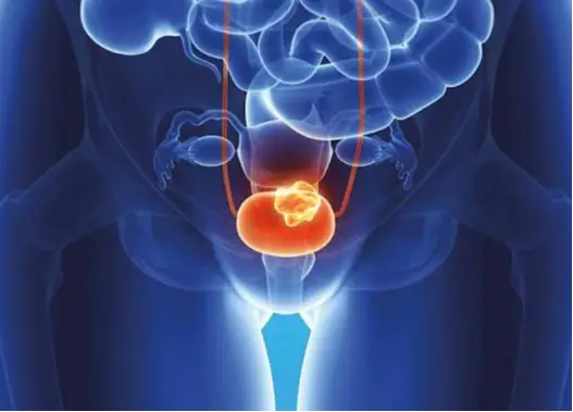
The bladder is an organ in the lower abdomen (pelvis) that can stretch to store urine and contract to expel it from the body. The bladder's main function is to store urine. Urine is produced by the bilateral kidneys and then transported to the bladder via two tubes called ureters. When you urinate, the muscles in the bladder contract, and urine exits the bladder at a certain rate through a tube called the urethra.
What is bladder cancer?
The bladder is an organ in the lower abdomen (pelvis) that can stretch to store urine and contract to expel it from the body. The bladder's main function is to store urine. Urine is produced by the bilateral kidneys and then transported to the bladder via two tubes called ureters. When you urinate, the muscles in the bladder contract, and urine exits the bladder at a certain rate through a tube called the urethra. Bladder cancer, or bladder tumor, can cause...malignant bladder cancerIt is an abnormal, uncontrolled tissue growth originating from the cells lining the inside of the bladder.Bladder tumor in menBladder cancer is 3-4 times more common in men than in women, which is partly attributed to higher rates of smoking and exposure to other carcinogens in men. Bladder cancer is the seventh most commonly diagnosed cancer in the male population worldwide and the most commonly diagnosed cancer type when both sexes are considered together. The incidence is 9 per 100,000 men and 2.2 per 100,000 women per year. It ranks second among urological cancers after prostate cancer. Worldwide, the mortality rate due to bladder cancer (per 100,000 people/year) is estimated at 3.2 for men and 0.9 for women.
Bladder cancer primarily affects older people. 90% of those diagnosed are over 50-55 years old. The average age of those diagnosed is 73. Bladder cancer is more common in people of African descent. At the initial diagnosis stage, 85% of patients present with the cancer confined to the bladder, not extending beyond it, while 15% have metastasized to lymph nodes or distant areas. Additionally, 70% of patients have superficial disease (non-muscle invasion). However, these patients have a 60-70% chance of recurrence and a 15-20% chance of progression (muscle spread) within two years. Bladder cancer has several stages. Treatment depends on the tumor's specific stage and grade, as well as the experience and skill of the medical team.
Types of Bladder Cancer - Pathology
Urothelial carcinoma - Transitional epithelial cancer:Urothelial carcinoma, also known as transitional cell carcinoma (TCC), is the most common type of bladder cancer (90-95%). In fact, if you have bladder cancer, it is almost certainly a urothelial carcinoma. These cancers originate from urothelial cells that line the inside of the bladder. Urothelial cells also line and cover the entire urinary tract, including the renal pelvis, ureters, and the portion where the urethra connects. In people with bladder cancer, the same type of cancer can sometimes develop in these areas simultaneously or at different times. Therefore, the entire urinary tract needs to be checked for tumors.
Squamous cell carcinoma:Only 1-3% of bladder cancers are squamous cell carcinomas. Under a microscope, the cells are very similar to the flat cells found in the skin. Almost all squamous cell carcinomas of the bladder generally have a more aggressive prognosis and are more likely to spread.
Adenocarcinoma:Before birth, there is a connection between the navel and the bladder. This is called the urachus. If part of this connection persists after birth, it can later develop into cancer. Cancers that originate in the urachus are a type of adenocarcinoma. Bladder adenocarcinomas can originate in the urachus or bladder, or be metastatic. Approximately one-third of bladder adenocarcinomas originate here. However, it is rare and accounts for about 1% of all bladder cancers. Almost all bladder adenocarcinomas have a poor prognosis and are prone to spreading..
Small cell carcinoma:Less than 1% of bladder cancers are small cell carcinomas. They originate from nerve-like cells called neuroendocrine cells. These cancers generally grow rapidly and usually require treatment with chemotherapy similar to that used for small cell carcinoma of the lung.
Mesenteric Sarcoma:Sarcomas begin in the muscle cells of the bladder, but they are very rare. Sarcomas are initially treated like transitional epithelial cancers, but if chemotherapy is needed, different protocols are required.
Spread of bladder cancer:The bladder wall has many layers. Each layer is composed of different cell types. The vast majority of bladder cancers begin in the innermost layer of the bladder, called the urothelium or transitional epithelium, and gradually grow outside the bladder, into other layers of the bladder wall, and/or into the bladder itself, reaching large volumes and stages. As the stage increases, treatment becomes more difficult. Over time, the cancer can grow outside the bladder and into nearby structures. Bladder cancer spreads through the lymphatic system to the prostate, seminal vesicles, rectum, peritoneum, and abdomen; through lymphatic spread to the external and deep obturator, external iliac, presacral, iliac common, paraaortic, paracaval, and interaortocaval lymph nodes; and through the bloodstream to the lungs, liver, bones, and adrenal glands.
Superficial and disseminated (invasive and non-invasive) bladder cancer:Bladder cancer is divided into two groups: superficial or non-invasive, meaning it has spread to the bladder muscle. If a papillary or flat tumor grows into the deeper layers of the bladder, i.e., the muscle layer, it is called invasive urothelial carcinoma.
This fundamental distinction is made as follows:
- Superficial bladder cancers (Non-invasive):These cancers are only in the inner layer of the cells (transitional epithelium). They have not grown into deeper layers. 70-75% of cases are in this group. Their likelihood of spreading or invading is lower than that of flat cancers (15-20%).
- Invasive bladder cancers:The cancer can spread and progress into deeper layers of the bladder wall. These cancers are more likely to spread to other areas of the body and are more difficult to treat.
Papillary and flat bladder cancer types:Bladder cancers are also divided into two subtypes, papillary and flat, depending on how they grow. Essentially, since 2015, bladder cancers have also been divided into two groups based on their genetic and molecular content or structure: luminal and basal. In pathology, immunohistochemical differentiation between luminal (GATA3 positive) and basal (KRT5/6 positive) can be made with 90% accuracy. In addition, there is also a pathological entity called carcinoma in situ.
1- Papillary-lunminal carcinomas:They grow as thin, finger-like, vegetal cauliflower-like projections from the inner surface of the bladder into the bladder lumen. Papillary-luminal tumors generally grow into the bladder lumen without extending into deeper bladder layers. These tumors are called superficial, or non-invasive, papillary cancers. Most very low-grade (slow-growing), non-invasive papillary cancers are called papillary urothelial neoplasms with low metastasis potential and tend to have a very good prognosis.
Papillary-luminal cancers originate from the middle and superficial layers of the urothelium, have a better prognosis than the basal type, generally contain estrogen receptors, and exhibit differences in genetic and molecular markers (FGFR3, ELF3, CDKN1A, TSCI mutations) compared to the basal type. They are also more sensitive to immunotherapy than the basal type.
2-Flat carcinomas (basal type):They do not grow towards the inner cavity of the bladder. They tend to spread from the inner surface of the bladder outwards, towards the muscle layer. Basal type bladder cancer is more often flat, originates from the bottom-basal layer of the urothelium, has a worse prognosis than the luminal type, shows differences in molecular and genetic markers (TP53, RBI mutations are prominent) compared to the basal type, and is more sensitive to cisplatin-based chemotherapy than the luminal type. The incidence of sarcomatoid and squamous cell transformation, considered a sign of poor prognosis in the variant type, is higher in the basal type compared to the luminal type. Basal type bladder cancer is more common in muscle-invasive cancers in women. This may be one reason for the poor prognosis of bladder cancer in women.
3-Carcinoma in-situ:If a flat tumor is located only in the inner layer of bladder cells and has not yet spread to the deeper layers, it is called carcinoma in situ (non-invasive flat carcinoma - CIS). CIS is a high-grade transitional epithelial cancer with mucosal boundaries and a velvety-grassy appearance. Its clinical significance lies in its potential to invade and spread in 40-60% of cases.
Carcinoma in situ is divided into 3 groups according to its clinical type.
- Primary: Isolated primary carcinoma in situ not associated with a papillary tumor;
- Secondary: Recurrence as carcinoma in situ in patients who previously had a papillary tumor.
- Simultaneous: Carcinoma in situ accompanying the presence of a urothelial tumor in the bladder.
Risk Factors for Bladder Cancer
While there is no single definitive cause of bladder cancer, numerous known risk factors are implicated, although the presence of these risk factors does not necessarily lead to cancer. Modifiable factors include smoking and exposure to carcinogens, while unmodifiable factors include age, genetics, and family history. Bladder cancer can also occur without any associated risk factors.
Tobacco and tobacco products (cigarettes):Smoking is one of the most important risk factors. The incidence of bladder cancer is 3-4 times higher in smokers. The number, dose, and duration of smoking all increase the risk. It is reported that the risk decreases by 30-60% within 2 to 4 years in those who quit smoking. Although it is not fully known how smoking affects the development of bladder cancer, it is thought to be due to carcinogenic substances such as aromatic amines and polycyclic aromatic hydrocarbons found in cigarettes. According to studies, tobacco contains many carcinogenic substances and is responsible for almost half of bladder cancer cases.
Occupational exposure and chemicals used:Occupational exposure to aromatic amines, polycyclic aromatic hydrocarbons, and chlorinated hydrocarbons accounts for approximately 10% of all cases and is the second most important risk factor for bladder cancer. Many occupational groups, such as paint industry workers, dry cleaners, textile workers, rubber and aluminum workers, chemical industry workers, painters, oil workers, dental technicians, and hairdressers, are at risk due to exposure to certain chemicals. Among these chemicals, substances proven to be carcinogenic (such as 2-Naphthylamine, 4-Aminobiphenyl, benzene, and benzidine) are thought to concentrate in urine, have a carcinogenic effect on the cells lining the bladder, and cause gene mutations in bladder cells.
Infection-inflammation:In areas where schistosomiasis (bilharziasis), a parasitic disease, is endemic, squamous cell bladder carcinoma develops in approximately 5% of infected cases. Similarly, long-term use of permanent urinary catheters and conditions causing chronic irritation such as bladder stones increase the risk of squamous cell carcinoma of the bladder. In patients with neurogenic bladder, it has been shown that this risk can be reduced by using condom catheters instead of permanent catheters or by performing clean intermittent catheterization..
Drugs-Radiotherapy:The painkiller phenacetin, the cancer drug cyclophosphamide, and a well-known diabetes medication (PGZN) are all drugs that carry a risk for bladder cancer. Food supplements containing aristolochic acid have also been shown to increase the risk of bladder cancer. Radiation therapy (radiation treatments) to the pelvic region, whether for gynecological cancers or any other reason, also poses a risk for bladder cancer. This exposure increases the risk of bladder cancer by 3-4 times compared to the general population.
Water Consumption and Diet:Increased water consumption reduces the risk of bladder cancer. This may be because the bladder empties more frequently, which is associated with carcinogenic substances remaining in the bladder for a shorter period and at lower concentrations. Chlorination of drinking water and increased trihalomethane levels are potentially carcinogenic, and exposure to high levels of arsenic in drinking water (arsenic-contaminated water) also increases the risk of bladder cancer. Smoking has a combined effect with arsenic exposure. While the relationship between the use of personal hair dye and risk remains unclear, an increased risk has been observed in individuals using permanent hair dye and those with slow NAT2-acetylation. The effect of metabolic factors (body mass index, blood pressure, plasma glucose, cholesterol, and triglycerides) is unclear. Dietary habits appear to have little effect; recently, flavonoids, which give plants bright colors such as yellow, orange, and red and perform antioxidant functions in the human body, have been found to have a protective effect. The Mediterranean diet, characterized by the consumption of vegetables and unsaturated fats (olive oil) and moderate protein intake, has been reported to be associated with a lower incidence of bladder cancer.
Age and Gender:The risk of bladder cancer increases with age. Approximately 90% of people with bladder cancer are over 55 years old. Bladder cancer is 3-4 times more common in men than in women.
Genetic and familial predisposition:Individuals with family members who have had bladder cancer are at a higher risk of developing the disease themselves. While family history appears to have little effect, and to date, no clear significance has been shown for any genetic variation in bladder cancer, the genetic events leading to bladder cancer are not fully understood. Genetic predisposition does have an influence on the risk of developing bladder cancer through the influence of other factors. However, multiple mechanisms are thought to play a role, such as the activation of oncogenes—genes that help cells grow, divide, and survive—and the inactivation of tumor suppressor genes, which normally control cell division, repair errors in DNA, or cause cells to die at the right time. Some of these include genetic deletions on chromosome 9, mutations in the p53 tumor suppressor gene, chromosome 17p deletion, and loss of chromosome 11p containing the c-Ha-ras proto-oncogene.
It is a known fact that a mutation in the retinoblastoma (RB1) gene increases the risk of bladder cancer. Cowden disease, caused by mutations in the PTEN gene, is primarily associated with breast and thyroid cancers. Individuals with this disease also have a higher risk of developing bladder cancer. Lynch syndrome is primarily associated with colon and endometrial cancer. Individuals with this syndrome have also been found to have an increased risk of bladder cancer.
Having bladder cancer:If a patient has a history of bladder cancer, the likelihood of developing the same type of cancer again in different parts of the bladder, or in the ureter and renal pelvis, is quite high. Therefore, patients with bladder cancer need careful monitoring to check different areas of their urinary tract.
Congenital bladder defects:Before birth, there is a connection between the navel and the bladder. This is called the urachus. If part of this connection persists after birth, it can develop into cancer later. Cancers that begin in the urachus are a type of adenocarcinoma. Approximately one-third of bladder adenocarcinomas originate here. However, it is rare and accounts for about 1% of all bladder cancers. Another rare congenital anomaly called exstrophy significantly increases the risk of bladder cancer. In bladder exstrophy, both the bladder and the abdominal wall in front of the bladder fail to close completely during development in the womb.
What are the symptoms of bladder cancer?
Presence of blood in the urine - Bloody urination (hematuria):Blood in the urine is the most common symptom of bladder cancer. It is usually painless and contains clots. Depending on the amount of blood in the urine, visible bleeding can range from pink to dark red, or the urine may be normal with only microscopic blood detected through urinalysis. The bleeding may not be continuous; it may be present one day and absent the next, and the urine may remain clear for weeks or even months. However, if bladder cancer is present, blood in the urine will reappear over time. In the early stages of bladder cancer, it usually causes bleeding and may not cause pain or other complaints. Sometimes, mild pain or burning during urination may occur along with bleeding. Blood in the urine does not always mean you have bladder cancer. It can be caused by urinary tract infections, kidney and prostate cancer, kidney, bladder, and urinary tract stones, prostate enlargement, and many other reasons other than bladder cancer.
Changes in urination habits and burning/dysuria symptoms:These symptoms do not only occur in bladder cancer; they can also be caused by urinary tract infections, kidney and prostate cancer, kidney, bladder and urinary tract stones, prostate enlargement, and many other conditions besides bladder cancer.
These are the complaints:
- Need to urinate more frequently than usual
- Pain or burning while urinating
- Urgent need to urinate even when the bladder is not full enough.
- Difficulty urinating or a weak urine stream
- The need to urinate multiple times throughout the night.
Symptoms of advanced bladder cancer:Large bladder cancers, or those that have spread to other parts of the body, can sometimes cause other symptoms such as those listed below. Again, many of these complaints could be caused by something other than bladder cancer, but it is important to have them checked.
Symptoms of advanced cancer may include:
- Inability to urinate
- Back pain on one side
- Loss of appetite and weight loss
- Feeling tired or weak
- Swelling in the feet
- Bone pain
How is bladder cancer diagnosed?
Bladder cancer is usually diagnosed primarily based on the patient's symptoms and complaints. Sometimes, bladder cancer can be detected during routine checkups. Beyond these, if bladder cancer is suspected, further examinations and tests are needed to confirm the diagnosis.
1- Medical history and physical examination:To learn more about your symptoms, information about your medical history and family history can be valuable in diagnosing bladder cancer.
2-Physical examination:A systematic physical examination is the first step in diagnosis, aiming to gather information about possible bladder cancer symptoms and other health problems. In addition, a digital rectal examination and, if necessary, a gynecological examination in women may be performed. During these examinations, it may sometimes be possible to palpate a bladder tumor, determine its size, and assess whether it has spread.
3- Urine analysis:The first step in diagnosing bladder cancer is a urinalysis, checking for blood in the urine (hematuria). Blood in the urine is often caused by non-cancerous problems such as infections, but it is also an early sign of bladder cancer. Blood in the urine may be microscopic or large enough to be visible to the naked eye. In some patients, a culture may be ordered to check for a urinary tract infection.
4- Urine cytology:This test involves searching for shed cancer cells in fresh urine or bladder cleansing fluid under a microscope. Urine cytology is important in diagnosing bladder cancer, but it is more helpful in identifying high-grade tumors; therefore, it is not sufficiently reliable as a screening test. A normal result does not necessarily mean there is no bladder cancer, and it is not a reliable enough test to guide diagnosis. Certain standards have been established for reporting this test results.
A standardized reporting system was published by the Paris Working Group in 2016.
- Sufficiency of urine samples;
- Negative for high-grade urothelial carcinoma (Negative);
- atypical urothelial cells
- Suspected for high-grade urothelial carcinoma (Suspect);
- high-grade urothelial carcinoma
- low-grade urothelial neoplasia
5- Urine tests for tumor markers:These tests can detect certain substances in urine that indicate bladder cancer, thus aiding in its diagnosis, but none of these tests are reliable enough for diagnosis, and regardless of the results, they do not provide an absolute answer for diagnosis and follow-up. They are more reliable than cytology. They may miss some bladder cancers, or in some cases, the test result may be abnormal even in people without cancer. Currently, these tests are mainly used to look for bladder cancer in people with symptoms and complaints, or as an aid in diagnosing recurrence in bladder cancer follow-up. Blood in the urine, the effect of BCG, and infections affect the results of these tests. According to current information, no urine marker can replace cystoscopy during follow-up or reduce the frequency of routine cystoscopy.
These tests:
- UroVysion ™(FISH): This test helps detect chromosomal changes that are common in bladder cancer cells.
- BTA tests - BTA - Stat and Trac:These tests detect a substance in urine called bladder tumor antigen (BTA), also known as human complement factor-related protein (CF-Hrp). There are two measurement methods: BTA-trac quantitative-ELISA and BTA-stat, which is a direct test similar to a pregnancy test.
- ImmunoCyt ™: This test is used to detect the presence of substances such as mucin and carcinoembryonic antigen (CEA), which are commonly found in cancer cells.
- NMP22 BladderChek®: This test looks for a protein called NMP22 (nuclear matrix protein 22) in the urine. People with bladder cancer often have higher levels of it.
- Fibroblast Growth Factor Receptor (FGFR), Telomerase Reverse Transcriptase (TERT), Microsatellite analysis, etc.
6-Cystoscopy:Bladder cancer is diagnosed through cystoscopy and biopsy. Cystoscopy is the most important and fundamental endoscopic method used in the diagnosis of bladder cancer and in monitoring recurrence after treatment. It allows examination of the bladder and urethra using a thin, camera-equipped, lighted tube called a cystoscope. Cystoscopy for bladder cancer diagnosis not only allows for diagnosis but also enables biopsy, collection of samples for lavage cytology, and, if necessary, surgical intervention for first-line treatment of bladder cancer, namely transurethral resection of bladder cancer (TUR-MT), all performed in the same session. This is what we call cystoscopy and the necessary procedure. A bimanual examination must be performed while the patient is under anesthesia. This allows for some insight into the extent and stage of the existing cancer. In cystoscopic imaging, lesions that appear as papillary lesions generally indicate a low-grade and non-invasive tumor, while solid and flat lesions suggest a higher-grade and more aggressive tumor. Furthermore, if complete removal of the tumor, including the muscle layer, is not possible (first-line treatment), a portion of the tumor, including the muscle layer, is removed and sent for pathology. Cystoscopy is generally performed with a rigid (hard-metal) cystoscope, but it can also be performed with a flexible (flexible) cystoscope, although these are not as convenient and efficient as rigid cystoscopes. New cystoscopy techniques have been developed to improve the effectiveness of cystoscopy.
These:
- Narrowband imaging:Narrowband imaging (NBI) involves applying specific blue and green wavelengths of light to the inner lining of the bladder during a standard cystoscopy procedure. This enhances the visual contrast between healthy and cancerous tissue, making it easier to detect tumors in the bladder. This method does not require instillation of contrast agents into the bladder.
- Floresans sistoskopi(Also known as ultraviolet light cystoscopy) This can be performed in conjunction with routine cystoscopy. For this examination, a light-activated medication is placed in the bladder during cystoscopy. It is taken up by cancer cells. When the doctor then shines a blue light through the cystoscope, any cells containing the medication will glow (fluorescent). This can help the doctor see abnormal areas that might have been missed by the white light normally used.
- Confocal laser micro-endoscopy:It is a high-resolution imaging probe designed to provide real-time endoscopic histological grading, but it is not yet in standard use; evidence regarding its usefulness is needed.
- SPIES imaging system:Storz's professional image enhancement system (IMAGE1 S, officially called SPIES) is an image enhancement system that uses four different light spectrums. Again, with this method, we don't yet have much evidence-based information regarding its results.
7-Transurethral resection of bladder tumor (TUR-MT):If any abnormal areas are seen during cystoscopy, biopsies are taken from those areas to determine if cancer is present, and if possible, the tumor is removed as a first-line treatment. Taking a biopsy from the abnormal area is the procedure used for first-line treatment if an obvious tumor is present: closed bladder tumor surgery, also known as transurethral resection of the bladder tumor (TUR-MT). During this procedure, the tumor and a portion of the bladder muscle surrounding the tumor are removed. The removed samples are then sent to a pathology laboratory. If cancer is found, the test determines whether it has invaded the muscle layer of the bladder wall (spread). This allows us to determine both whether cancer is present and, if so, whether it has spread to the muscle layer. These results provide invaluable contributions to tumor staging. Bladder cancer can sometimes develop in more than one area of the bladder (or other parts of the urinary tract). Therefore, biopsy samples may be taken from several different locations in the bladder and urinary tract, especially if cancer is strongly suspected but no obvious tumor is visible. The bladder lavage fluid obtained is also sent to pathology for cytological analysis.
8- Biopsy results:Biopsy samples are examined in a laboratory by a pathologist and sent to another laboratory for testing. If bladder cancer is detected, the extent of tumor spread, cell type, and cancer grade are determined. The degree of invasiveness in the biopsy can indicate how deeply the cancer has grown into the bladder wall. This is crucial in deciding on treatment.
Accordingly:
- Non-invasive bladder cancer:Cancer is called non-invasive if it remains within the inner layer of cells without growing into deeper layers. This term can also be used to describe superficial or non-invasive bladder cancer.
- Invasive bladder cancer:If cancer grows into deeper layers of the bladder, it is called invasive. Invasive cancers are more likely to spread and are more difficult to treat.
Cancer Cell Grading:Bladder cancers are graded based on how malignant the cancer cells appear under a microscope. Low-grade cancers resemble normal bladder tissue and are often called well-differentiated cancers. These types of cancers generally have a good prognosis. High-grade cancers are less similar to normal tissue and are more aggressive. These cancers can also be described as poorly differentiated or undifferentiated. High-grade cancers are more likely to grow into the bladder wall and spread outside the bladder. These cancers are more difficult to treat.
9- Intravenous pyelography (IVP):Intravenous pyelography (IVP), also called intravenous urography (IVU), is an X-ray of the entire urinary system taken after a special dye is injected into a vein. This dye is removed from the bloodstream by the kidneys and then passes into the ureters and bladder. X-rays are taken as this happens. The dye outlines these organs on the X-ray and helps to show urinary tract tumors. Patients with a history of allergies or any type of kidney problem may have another test chosen instead. IVP is not routinely used for diagnosing bladder cancer; computed tomography (CT) scans can provide more valuable information.
Retrograde pyelogram (RPG):For this test, a catheter (thin tube) is inserted into the bladder or a ureter via the urethra. A contrast agent is then injected through this catheter, and the bladder, ureters, and renal cavities are imaged using X-rays. This is not among the most commonly used methods, unlike IVP (intravenous pyelography).
10-Computed tomography (CT) scan:A CT scan uses X-rays to obtain detailed cross-sectional images of your body. A computed tomographic examination of the kidneys, ureters, and bladder is called CT-urography. It can provide detailed information about the size, shape, and location of any tumors in the urinary tract, including the bladder. It can also help show enlarged lymph nodes in the abdomen and pelvis that may be cancerous, as well as other organs, and determine if there are metastases.
CT-guided needle biopsy:CT scans can also be used to guide a biopsy needle to a suspicious tumor. This is used to obtain samples from areas where the cancer may have spread.
11-Magnetic resonance imaging (MRI) scan:Like CT scans, MRI scans provide detailed images of soft tissues in the body. However, MRI scans use radio waves and powerful magnets instead of x-rays. MRI imaging gives us an idea of whether bladder cancer has spread to neighboring tissues and organs, or to nearby tissues or lymph nodes. MRI-urography, a specialized method for imaging the upper part of the urinary system (ureter and renal pelvis), can be performed.
Multiparametric magnetic resonance imaging (mpMRI)Its role in the diagnosis and staging of bladder cancer has not yet been determined. A standardized mpMRI reporting methodology for patients with bladder cancer (VIRADS) was recently published but has not yet become standard practice.
12-Ultrasound: UUltrasound can be useful in determining the size of bladder cancer and whether it has spread outside the bladder to nearby organs or tissues. It can also be used to check the kidneys. It is an easy and simple method.
Ultrasound-guided needle biopsy: Ultrasound can also be used to guide a biopsy of a suspected cancerous area in the abdomen or pelvis.
13-Chest X-ray:A chest X-ray can be taken to see if bladder cancer has spread to the lungs. However, this is not commonly preferred today as chest CT scans are usually used for this purpose.
14-Bone scan:Bone scans can help look for cancer that has spread to the bones. This test is usually done if you don't have symptoms like bone pain or if blood tests show that cancer may have spread to your bones. For this, a small amount of a low-level radioactive substance is injected into the damaged bone areas. A special camera counts the radioactivity intensity, and the entire skeletal system can be visualized. A bone scan can show cancer in the bone, but to be sure, it may need to be confirmed with other imaging methods such as plain X-rays, MRI scans, or even a bone biopsy.
15- Positron emission tomography (PET) scan:It is a combination of radioactive material and CT scan (PET/CT) and is increasingly used to detect the spread of bladder cancer to lymph nodes and other organs. PET-CT can increase the likelihood of finding distant metastases. However, it is not highly recommended for staging bladder tumors.
Staging and Grading of Bladder Cancer
After a bladder cancer diagnosis, studies are conducted to determine whether the cancer has spread and, if so, how far. This process is called staging. The stage of a cancer describes the size and extent of the cancer in the body. Staging helps determine how serious the cancer is and how best to treat it. The stage is one of the most important factors in predicting how the cancer will be treated and how successful the treatment will be. The staging of bladder cancer is based on the results of physical examinations, biopsies, and imaging tests (CT or MRI scans, X-rays, etc.) mentioned in the bladder cancer diagnosis, as well as the results of surgery.
To determine the stage of cancer, the following questions are answered:
- How deep has the cancer progressed into the bladder wall, and if so, to what extent?
- Has the cancer spread to organs and tissues adjacent to the bladder?
- Has the cancer spread to lymph nodes adjacent to the bladder or to distant organs?
Tumor, Lymph Node, Metastasis Classification (TNM):The 2009 TNM classification, approved by the Union International Contre le Cancer (UICC), was updated in 2017, but no changes were made regarding bladder tumors. It is based on 3 fundamental pieces of information.
- T: It describes how far the main (primary) tumor has grown along the bladder wall and whether it has spread to nearby tissues.
- N: This indicates cancer that has spread to lymph nodes near the bladder. Lymph nodes are bean-sized collections of immune system cells where cancers often spread first.
- M: It shows whether the cancer has spread (metastasis) to other organs such as the lungs or liver, or to distant areas such as lymph nodes not near the bladder.
- Numbers or letters after T, N, and M,This provides more details about each of these factors. Higher numbers mean the cancer is more advanced. Once a person's T, N, and M categories are determined, a final stage is usually determined after surgery.
As a rule, the lower the number, the less the cancer has spread. A higher number, such as stage IV, means a more advanced cancer. And in a stage, the earlier letter indicates a lower stage. Cancers with similar stages tend to have a similar appearance and are generally treated similarly.
The staging system in the table below refers to definitive pathological staging. It is based on physical examination, biopsy, imaging test results, and postoperative pathological data. This staging is more precise than clinical staging, which is determined solely based on preoperative analysis data.
TNM Staging for Bladder Cancer (2017)
T - Tumor:
TX: Tumor cannot be evaluated.
T0: No evidence of tumor presence.
Ta: Non-invasive - Non-spreading papillary carcinoma
Tis: Carcinoma In-sutu: "flat tumor"
T1: Tumor has invaded and spread to the subepithelial connective tissue.
T2: Tumor has spread to the muscle layer.
T2a: The tumor has spread to half of the muscle layer.
T2b: The tumor has spread into the deep muscle layer, meaning it has spread beyond the muscle's innermost layer.
T3: The tumor has spread outside the bladder and into the perivesical tissue.
T3a: Microscopically leaked outside the bladder.
T3b: Macroscopically visible extravesical mass (mass extending outside the bladder).
T4: The tumor has spread to any of the following: prostate stroma, seminal vesicles,
uterus, vagina, pelvic wall, abdominal wall
T4a: The tumor has spread to the prostate stroma, seminal vesicles, uterus, or vagina.
T4b: The tumor has spread to the pelvic wall or abdominal wall.
N - Regional lymph nodes
NX: Regional lymph nodes cannot be evaluated.
N0: No regional lymph node metastasis.
N1: Metastasis in a single lymph node in the pelvis (hypogastric, obturator, external iliac or
presacral)
N2: Metastasis in multiple regional lymph nodes in the true pelvis (hypogastric, obturator,
external iliac or presacral)
N3: Extensive metastasis in iliac lymph nodes
M - Distant metastasis
M0: No distant metastases
M1a: Metastasis to non-regional lymph nodes
M1b: Metastasis to other distant areas and organs.
Subclassification of T1:It has been reported that the subcategorization of T1 can significantly contribute to determining the course and treatment of cancer. The depth and extent of the lamina propria have been shown to have prognostic value. Its use is most recently recommended according to the World Health Organization (WHO) classification from 2016.
Histological grading of bladder urothelial carcinomas:
1973 WHO rating
- 1st degree: well-differentiated
- 2nd Degree: Moderately differentiated
- Grade 3: poorly differentiated
2004/2016 WHO grading system (papillary lesions)
- Papillary urothelial neoplasm with low malignant potential (PUNLMP)
- Low-grade (LG) papillary urothelial carcinoma
- High-grade (HG) papillary urothelial carcinoma
SO 2004 Histological classification for flat lesions
1- For non-malignant lesions:
- Urothelial proliferation of unknown malignant potential (flat lesion without atypia or papillary aspects).
- Reactive atypia (atypical flat lesion).
- Atypia of unknown significance.
- Urothelial dysplasia.
2. For malignant lesions: Urothelial CIS is always high grade.
Variant types of urothelial carcinomas and lymphovascular invasion: Micropapillary, plasmacytoid, and sarcomatoid urothelial carcinoma variants have an even worse prognosis than pure high-grade urothelial carcinoma. The presence of lymphovascular invasion (LVI) on pathological examination is a factor that increases the pathological staging grade and is associated with a worse prognosis.
Variant Types:
- Urothelial carcinoma (more than 90% of all cases);
- Urothelial carcinomas with partial squamous and/or glandular or trophoblastic differentiation;
- Micropapillary urothelial carcinoma;
- Nested variant and microcystic urothelial carcinoma;
- Plasmocytoid, giant cell, signet ring, scattered, undifferentiated;
- Lymphoepithelioma-like;
- Some urothelial carcinomas, along with other rare differentiations; small cell carcinomas; sarcomatoid urothelial carcinoma.
Molecular classification:Molecular markers and their prognostic role have been investigated. These methods, particularly complex approaches such as classifying patients based on molecular classification, are promising, but are not yet suitable for routine application.
Classification of Bladder Cancer Patients into Risk Groups
- Low-risk tumors:Primary tumor, single lesion, Ta, G1 (low-grade), less than 3cm, no concomitant CIS.
- Intermediate risk tumors:It includes all superficial tumors between low and high risk. Patients with annual or no recurrence, and those who received intracavitary chemotherapy immediately after TUR-MT are included in this coverage.
- High-risk tumors:All stage T1 tumors, all high-grade tumors (grade 3), CIS, and multiple tumors with frequent recurrence (more than once a year), tumors larger than 3cm, and stage Ta-low/intermediate grade (grade 1-2) tumors are included in this scope.
- The subgroup of highest-risk tumors:This includes stage T1 and high-grade (grade 3) tumors accompanied by CIS lesions, numerous and/or large-volume and/or recurrent T1 and high-grade tumors, T1 and high-grade tumors in the prostatic urethra accompanied by CIS, certain variant histological forms of urothelial carcinoma, and tumors showing lymphovascular invasion.
Survival Rates for Bladder Cancer
Survival rates are figures that give an idea of what percentage of people with the same type and stage of cancer are still alive for a certain period of time (usually 5 years) after diagnosis. They can't tell you exactly how long you'll live, but they can help you better understand how likely your treatment is to be successful. It's important to remember that survival rates are estimates and are often based on previous outcomes for a large number of people with a particular cancer, but they cannot predict what will happen in any given person's case. These statistics can be confusing and may raise further questions.
These groups are formed as follows:
- Localized disease:There is no indication that the cancer has spread outside the bladder.
- Regional spread:The cancer has spread from the bladder to nearby structures or lymph nodes.
- Distant metastasis:Cancer has spread to distant parts of the body, such as the lungs, liver, or bones.
Five-year survival rates for bladder cancer.
- Carcinoma in situ alone: 96%
- Localized disease: 69%
- Regional spread: 37%
- Distant metastasis: 6%
- If all stages are combined: 77%
Bladder Cancer Treatment, Treatment of Bladder Cancer, Bladder Tumor Treatment
A multidisciplinary approach to bladder cancer treatment.
Decision-making in cancer treatment requires combining a large amount of data, including symptoms and imaging information such as MRI/CT scans and PET scans, and necessitates a multidisciplinary approach—integration of all relevant departments—to select the most appropriate treatment for the patient. International standards in cancer treatment require this approach.
At Prof. Dr. Hakkı Perk's private clinic, treatment planning for uro-oncological, or cancer, patients is carried out using a multidisciplinary approach. Once a week, at a hospital with which I have an agreement, cancer patients are discussed in an oncological council attended by urologists, medical oncologists, radiation oncology specialists, nuclear medicine specialists, pathologists, and radiologists. Treatment decisions and processes for these patients are finalized. Treatment processes for patients with prostate cancer, kidney cancer, adrenal gland tumors, bladder cancer, and testicular cancer are determined with the participation of all relevant departments.
Furthermore, cancer patients are guided by international oncology guidelines such as NCCN, ASCO, EAU, and AUA in their treatment management. In such multidisciplinary collaborations and cancer councils, all aspects of a patient's condition are considered and decided upon. Feedback presentations regarding patients undergoing treatment are also given at these councils.
A- Bladder Cancer Surgery
1-Transurethral resection of bladder tumor (TUR-MT), Bladder TUR surgery
Transurethral resection of a bladder tumor (TUR-MT) is the first and most important step in bladder cancer treatment. The goal of TUR-MT is to accurately diagnose and completely remove all visible tumors. This surgery is crucial for pathological diagnosis, assessment of the extent of cancer, and treatment planning, and may be required repeatedly. TUR-MT is also the most common treatment method for early-stage or superficial (pt1 bladder cancer, i.e., non-muscle invasive - Ta, T1, and Carcinoma in Situ) bladder cancers. 70-75% of bladder cancers are superficial at initial presentation, and TUR-MT is the first-line treatment for these. The aim of TUR-MT is to remove cancer cells and surrounding tissues down to the muscle layer of the bladder wall.
TUR-MT (Re-TUR) again:In some cases, it may become necessary to repeat this surgical procedure 2-4 weeks after the initial operation. These necessities include: the procedure not being performed properly or the tumor not being completely removed during TUR-MT; the absence of muscle tissue in the tissue removed during TUR-MT, meaning insufficient basal tissue was taken; and Re-TUR-MT is necessary for all T1-stage tumors and all high-grade tumors. If the tumor is low-grade Ta or primary carcinoma in situ (CIS), re-TUR is rarely needed.
How to do TUR-MT?:This is a closed endoscopic surgery performed under general or local anesthesia, accessed through the urethra. A thin, rigid cystoscope called a resectoscope is inserted into your bladder via your urethra. The tip of the resectoscope has a wire loop (1c diameter, which can also be used to measure the tumor diameter) used to remove tissue or tumors. Energy (monopolar or bipolar cautery, laser, etc.) is applied through this loop, and the removed tissues are sent to a pathology clinic for analysis. Tumors can be removed en bloc (total) or fractionally. An important criterion is taking a biopsy from the tumor base and the surrounding muscle layer; this is crucial for tumor staging and determining the quality of the resection. Sampling at the tumor-normal tissue boundary is also beneficial. The cleaned areas are cauterized (fulguration) using a reusable energy system, which both controls bleeding and helps destroy any remaining cancer cells.
The side effects of TUR-MT are generally mild and usually do not last long. There may be some bleeding and pain when urinating immediately after the procedure. You can usually go home the same day or the next day and return to your normal daily life within one or two weeks.
Even if the tumor is completely removed with TUR-MT, bladder cancer has a high chance of recurring within 2 years, so the procedure often needs to be repeated. Frequent repetition of this procedure can lead to some loss of bladder function in some patients, resulting in urinary incontinence, decreased bladder capacity, and some impairment of bladder function in the long term.
In patients with a history of long-term recurrence of non-invasive, superficial, low-grade tumors, the surgeon may use fulguration to cauterize (instead of removing) small tumors seen during cystoscopy. This procedure can be performed in the office under local anesthesia. It is safe, but can be somewhat uncomfortable and is not highly recommended.
2-Cystectomy
Bladder cancer is muscle-invasive (spread) (Stage 2-4), meaning...stage 2 bladder cancerIn advanced stages of bladder cancer, removal of all or part of the bladder may be necessary. This operation is called a cystectomy. Often, chemotherapy is given before the cystectomy, but in some cases it may be given after.
Partial cystectomy:If cancer has progressed into the muscle layer of the bladder wall but is not very large and is in a single area, it may sometimes be appropriate to remove only the cancerous part of the bladder instead of removing the entire bladder. Lymph nodes adjacent to the bladder are also removed, which is important for staging the disease. However, the percentage of patients with metastatic bladder cancer suitable for this procedure is quite low (10%). The main advantage of this surgery is that it preserves the patient's bladder and avoids the need for much larger surgery that would profoundly affect their quality of life. However, it can reduce the remaining bladder capacity, meaning they will need to urinate more frequently. The main concern with this type of surgery is the high probability of bladder cancer recurring in another part of the bladder wall. Therefore, this procedure is recommended only for very select patient groups.
Radical cystectomy, which is the removal of the bladder in bladder cancer:In fact, the primary treatment for invasive bladder cancer that has spread to the muscle is reconstructive surgery called radical cystectomy and urinary diversion. In this operation, the entire bladder and adjacent lymph nodes are completely removed. In men, the prostate and seminal vesicles are also removed. In women, the ovaries, fallopian tubes (the tubes connecting the ovaries and uterus), uterus, cervix, and a small portion of the vagina are also removed.
Most often, a cystectomy is performed through an incision in the abdomen. You will need to stay in the hospital for about a week after the surgery. You can usually return to your normal life a few weeks later.
Radical cystectomy and subsequent reconstructive surgery can be performed via open surgery, closed laparoscopic surgery, or robotic surgery. The choice of surgical method depends on the knowledge and experience of the surgical team. Closed laparoscopic or robotic surgery results in less pain and faster postoperative recovery due to smaller incisions. However, long-term data to compare results with standard open surgery is lacking. Therefore, its effectiveness is not yet entirely clear. It is crucial that any type of cystectomy is performed by a surgeon experienced in bladder cancer treatment. If the surgery is not performed properly, the likelihood of cancer recurrence is higher.
3-Reconstructive surgery after radical cystectomy (Urinary Diversion)
After the entire bladder is removed, a different pathway is needed to store and drain urine when needed, and this is called urinary diversion. Diversion can be performed using various techniques. Generally, we can divide diversions into two groups: incontinent (uninterrupted flow of urine collected outside the body) and continent (urine is stored in the body and drained when needed). When deciding on one of these techniques, the patient's kidney and liver function, mental state and compliance, expected lifespan, and the location and extent of the cancer are considered. The condition of the digestive system (intestines) is also a determining factor in choosing the technique to be applied. To determine the most suitable technique for patients, the limitations and side effects of each option should be thoroughly explained to the patients. In addition to personal preference, the patient's physical and mental ability to use and manage the diversion, and the presence or absence of social and family support are important factors in the decision.
In cases where the cancer is too widespread to be surgically removed (a condition known as frozen pelvis), and has invaded surrounding tissues and organs, a reconstructive procedure can be performed without removing the bladder. This involves separating the ureters and redirecting urine flow. In this case, the goal of the surgery is not to cure the cancer, but rather to protect the kidneys and prevent or alleviate obstruction of urine flow.
Urinary Diversion Techniques - Types of Reconstructive Surgery:
-Ileal loop (Bricker surgery):The most common method involves taking a short section of the small intestine (preferably the ileum, 10-15 cm), connecting the ureters (the tubes carrying urine from the kidneys) to this removed section of intestine, and then creating an anastomosis (urostomy) at one end of the removed section to the abdominal wall. This is called Bricker surgery or ileal loop. This creates a passageway known as the ileal loop for urine to pass from the kidneys to the outside of the body. Urine flows from the kidneys through the ureters into the ileal loop. After this procedure, a bag called a urostomy bag is attached to the skin of your abdomen around the stoma to collect the urine. Urine continuously fills this bag and is emptied as needed. This is called incontinence diversion because the flow of urine in your body cannot be controlled. This 'enteric stoma' creates more distance between the kidneys and the skin and generally reduces the likelihood of infection entering the body from the outside environment. Another advantage for patients is that this stoma is easier to manage and carries a smaller risk of stricture. This method is technically relatively simple and reliable, and therefore is the most commonly used form of diversion. When choosing this type of urinary diversion, it is important to be aware of the time and effort required to adapt to living with a bladder attached to the abdominal wall. Long-term complications such as recurrent infections, narrowing/scarring at the junction sites, urinary leakage or seepage, and urinary stone formation (urolithiasis) can occur after this type of surgery.
-Creation of a reservoir inside the body - Mainz pouch surgery:A section of the small and/or large intestine (sometimes using the appendix) is taken to create a reservoir in the abdominal cavity. This section is shaped like a bladder and then connected to the skin via a valve mechanism, also from the intestine. This low-pressure reservoir allows urine to accumulate in the body as if it were a bladder. The aim of this technique is to ensure continence and control reflux (backflow to the kidneys). The reservoir can be emptied with a small plastic catheter through intermittent catheterization every 2-6 hours. The opening can be placed anywhere in the lower abdomen or at the navel (umbilicus).
-Artificial Bladder-Creation of a new bladder from the small intestine (orthopedic neobladder):In this technique, a section of the small intestine (preferably the ileum, 40-45 cm) is harvested, shaped to resemble a bladder, and a new bladder is constructed. The ureters are attached to either side of the newly created bladder, and its lower end is connected to the urethra. The new bladder is then placed back in its original location. The patient retains control over urination as before, eliminating the need to carry an external bag. This technique results in a continent reservoir with a volume comparable to the original bladder.
With this artificial reservoir, you will not feel the urge to urinate as you would with your original bladder, so you should empty your bladder every 2-4 hours. The new artificial bladder is emptied by contracting the abdominal muscles and relaxing the pelvic floor muscles (Valsalva maneuver), in addition to applying pressure to the pelvic floor manually. Applying pressure to the pelvic floor with both hands helps to completely empty the new bladder. Intermittent catheterization is needed in ¼ of women to completely empty the new bladder, while it is generally not necessary in men. Therefore, factors such as normal liver and kidney function, a relatively long life expectancy, good mental state, and a functional pelvic floor are preferred for this type of surgery. This technique may not be suitable if the patient has undergone extensive abdominal surgery or received high-dose radiotherapy. The patient's good adaptation to this type of operation, their knowledge and understanding of the procedure's requirements, improves their quality of daily life and positively affects the results. The newly created reservoir needs time to settle and begin functioning. You will need to train your new bladder to increase its capacity, and a healthcare team should provide bladder training. Exercise is necessary to increase the capacity of the new bladder. Initially, urinary incontinence and a feeling of bloating may occur. Lifestyle changes and getting used to some daily routines (urinating at specific intervals) are important. You will not have the same urge to urinate as before, so you need a program provided by a healthcare team. Over time, most patients regain the ability to urinate normally during the day, but nighttime urinary incontinence may cause some problems.
Risks and side effects (complications) of cystectomy and urinary diversion surgery:
1- Possible problems that may occur during and immediately after surgery are as follows:
- Risks and problems related to anesthesia.
- August
- Risk of embolism and blood clots traveling to the legs or lungs.
- Running injuries to organs
- Infection
- Pain after surgery can be controlled with painkillers.
2. Effects of cystectomy on urination:Some physical changes resulting from bladder removal and urostomy can negatively impact your quality of life. In patients who have undergone radical cystectomy, reconstructive surgery to drain urine can present with certain problems depending on the technique used. Depending on the type of reconstruction, you may need to learn how to empty the urostomy bag or how to insert a catheter if a stoma (reservoir) has been created. In addition to these changes, urinary diversion and urostomy can also lead to:
- Infections
- Urine leaks
- incontinence
- Stone formation in the reservoir
- Obstruction of urine flow, narrowing problems
- Absorption problems (depending on the amount of bowel involved)
3. Effects of radical cystectomy on sexuality in men:In radical cystectomy, the prostate and seminal vesicles are removed along with the bladder. Since these glands produce most of the seminal fluid, removing them means a man will no longer ejaculate. If an orgasm occurs, it will be a dry orgasm, with no ejaculation. Due to nerve damage during surgery, many male patients experience impaired erectile function. This erectile dysfunction may improve somewhat over time in some patients. The younger the patient, the higher the chance of regaining the ability to achieve a full erection. This should be discussed with the healthcare team before surgery. New surgical techniques may offer some improvements in reducing the likelihood of erectile problems.
4. Sexual effects of radical cystectomy in women:This surgery removes the uterus, ovaries, and usually the upper/front ⅓ of the vagina. This can often make sex more uncomfortable for women. In this case, a procedure to reconstruct the vagina may be required (vaginal reconstruction). Whether or not you have reconstruction, there are many ways to make sex more comfortable. During radical cystectomy, if the nerve bundles running along either side of the vagina are damaged, it can affect the ability to orgasm. In cases where the entire urethra needs to be removed along with the bladder, blood flow to the clitoris can be affected, which can also affect sexual arousal.
5- Sexual effects of urostomy:It's normal for both men and women to be concerned about having sex with a urostomy. Ensuring the urostomy bag is properly positioned, fits correctly, and emptied before sex reduces the likelihood of urinary leakage. Choosing sexual positions that prevent your partner's weight from rubbing against the bag can also be important.
B- Intravesical Treatment for Bladder Cancer - Medicated Bladder Washing
Non-metastatic, superficial tumors (Stage Ta and T1 and Carcinoma in situ) removed by closed endoscopic TUR-MT may frequently recur over time (50-70%) and/or become invasive (muscle-spreading cancer) (15-20%), depending on the tumor grade. Therefore, it is beneficial to group patients exhibiting similar biological behavior together to personalize and standardize follow-up and treatment as much as possible, and efforts in this direction have been ongoing for a long time. An appropriate classification should aim to protect the patient from unnecessary treatments and interventions while preventing or reducing possible recurrences, and especially to protect against or minimize disease progression and death.
Intravesical instillation therapy (bladder irrigation) after TUR-MT is a treatment method used to reduce the likelihood of recurrence and progression, i.e., the development of an invasive character, and is performed in almost all patients. Intravesical chemotherapy or immunotherapy is used for this purpose. Intravesical applications are performed to prevent cancer recurrence, reduce the frequency of tumor recurrence, decrease the frequency of cystoscopies performed for control purposes, and improve the patient's quality of life. Furthermore, it can contribute to preventing progression (cancer becoming invasive); meanwhile, BCG (immunotherapy) is the only drug proven to contribute to progression prevention. The procedure involves irrigating the bladder with appropriate immunotherapy or chemotherapeutic drugs once a week for 4-6 weeks, 2-4 weeks after surgery. In intravesical treatment, the drugs are administered directly into the bladder via a catheter, the patient holds the drug in the bladder for 2 hours, then empties it into the toilet. This can be repeated for another 6 weeks if necessary. This is called induction/initial treatment. After a 4-6 week interval, in some cases, maintenance washes may be required for at least 1-2 years, with 3 doses per 3-week period every 3 months.
-Low/moderately non-invasive, non-metastatic bladder cancersIntravesical chemotherapy and immunotherapy (BCG) can be used. Some studies show that immunotherapy yields the best results. For low-grade Ta tumors, intravesical chemotherapy given immediately after surgery may be sufficient, and additional intravesical treatment may not be necessary.
-High-risk non-invasive superficial,Immunotherapy is preferred in rapidly growing (high-grade) bladder cancer with more than two lesions.Bladder cancer bcgTherefore, if there is a good response to induction therapy with BCG instillation, it is recommended to continue this with 3 years of maintenance therapy. Maintenance therapy, or intravesical immunotherapy treatment programs, vary. Maintenance therapy can be given monthly, every 3 months, or every 6 months for 3-6 weeks/dose; these time intervals and dose numbers may vary depending on the patient's response, and sometimes inter-clinic differences and different protocols may be applied.
Much higher stage (Stage II-III),In high-grade tumors, a dose of intravesical chemotherapy is administered within the first 24 hours after TUR-MT. If radical surgery is not possible, in Stage II and very rarely in Stage III, intravesical immunotherapy protocol can be applied after systemic chemotherapy or radiotherapy, even if the benefits are questionable.
A single dose of intravesical chemotherapy immediately following TUR-MT:If the tumor is completely and safely removed with TUR-MT and there are no signs of invasive growth, a single dose of intravesical chemotherapy should be instilled immediately after surgery (within the first few hours). This is the rule and standard practice. This single-dose instillation is not performed if there is a possibility of bladder perforation during resection or if postoperative bleeding is very severe. The single-dose instillation destroys tumor cells floating in the bladder after TUR-MT and also eliminates small residual tumor cells that may have been missed in the resection area. It may be sufficient for low-grade tumors, but it may not be sufficient for moderate-grade tumors, and additional doses may be needed. This procedure reduces the risk of disease recurrence. The medication is administered through a catheter inserted after surgery; the catheter is blocked and then opened after 2 hours.
There are two types of drugs used in intravesical treatment.
- Immunotherapy (BCG)
- Chemotherapy(MTX-C-C, Epirubicin, Gemstabine vs)
Intravesical immunotherapy (BCG): IImmunotherapy causes the body's own immune system to attack cancer cells. The tuberculosis vaccine, Bacillus Calmette-Guerin (BCG), is the most commonly used intravesical immunotherapy drug in the treatment of superficial, early-stage bladder cancer. It is used to help prevent cancer progression—from becoming invasive—and to prevent frequent recurrence. BCG is a vaccine form of the bacteria that causes tuberculosis; it is a live vaccine and generally does not cause serious illness. Nearly 20 subtypes of the tuberculosis bacteria have been identified, and none is superior to the others. BCG is administered into the bladder via a catheter. It reaches the cancer cells and stimulates and activates the immune system. Immune system cells then attack the bladder and target the bladder cancer cells. For BCG to work, it needs to come into contact with the cancer cells. Therefore, it is used as an intravesical treatment.
BCG side effects: Serious side effects occur in 5% of cases. In intracavitary treatment with BCG, flu-like symptoms such as fever, pain, chills, and fatigue are commonly observed. These may last for 2 to 3 days after treatment. It can also cause burning in the bladder, frequent urination, and even blood in the urine. Due to side effects, the rate of treatment discontinuation is not low in some patients. Its use is not recommended in patients with compromised immune systems, and it is also not preferred in patients over 70 years of age. Rarely, BCG can spread into the bloodstream and body, leading to a serious tuberculosis infection (1%). This can occur even years after treatment.
Categories of unsuccessful intravesical BCG treatment: In these cases, combinations of drugs other than BCG or in combination with BCG are recommended, or radical cystectomy becomes unavoidable.
- Tumor resistant to intravesical BCG
- Tumor recurrence despite intravesical BCG
- BCG-unresponsive tumor
- BCG intolerance (discontinuing BCG treatment due to side effects)
İntravezikal chemoterapi:This treatment involves administering an appropriate chemotherapy drug into the bladder via a catheter once a week for 4-6 weeks after surgery, for low-to-moderate tumors, and then draining it after a certain period. These drugs can directly destroy actively growing cancer cells. Intravesical chemotherapy has been found to reduce the annual recurrence rate by 40-50% in low-to-moderate risk patients. In some patients, three doses of maintenance intravesical chemotherapy are continued every three months for a year to prevent cancer recurrence. The length and frequency of repeated intravesical chemotherapy applications are still debated; however, it should not exceed one year. Some intravesical chemotherapy drugs can also be used in cases where immunotherapy has been ineffective.
The chemotherapy drugs used for this purpose are as follows:
- MTX-C-C (MMC) is the most commonly used drug for intravesical chemotherapy.
- Gemcitabine may cause fewer side effects than MTX and is less likely to be absorbed into the bloodstream.
- Epirubicin is also used for this purpose and its effects are no different from other drugs.
- Apaziquone is a new agent, an MTX-C derivative, used intracavitarily. The enzyme deoxymidine diaphorase plays a key role in activating this normally inactive drug. While this enzyme is absent from normal bladder tissue, it is present in high concentrations in 40% of bladder tumors. This suggests that apaziquone may exhibit selective toxicity against tumor cells.
- Valrubicin may be considered more often in cases unresponsive to BCG, but there is no consensus on this matter.
- Paclitaxel: a newly introduced product.
- C60070, vicinium, and MCC (Mycobacterial cellular complex). Not yet available for clinical use.
İThe main side effects of intravesical chemotherapy are bladder irritation and burning sensation, and blood in the urine. The biggest advantage of delivering the chemotherapeutic drug directly into the bladder is that the drugs generally do not reach or affect other parts of the body outside the bladder. Thus, side effects from these drugs are avoided.
New treatment options for patients who do not respond to standard intracavitary therapies.
Intracavitary BCG therapy after TUR-MT is currently the most effective treatment method for high-risk bladder tumors without muscle metastasis. The course of action for patients unresponsive to BCG has always been a subject of debate. Sometimes, a delay in radical cystectomy can lead to muscle-invasive disease. However, secondary intravesical therapies have a place in selected patients. Especially in recent years, adjuvant therapies that enhance the effectiveness of intracavitary treatment have been introduced. Therefore, while preserving the possibility of cystectomy and avoiding delay, secondary therapies can be considered in this high-risk group of patients with superficial bladder cancer.
These secondary intracavitary treatments include the following:
- BCG + interferon is another immunotherapy. IFN is an older immunomodulator. A combination of IFN + BCG at doses of 50-100 million units has been found to achieve a response of nearly 40% in patients who did not respond to BCG alone; however, this response lasted for more than two years in only one-third of these patients. This agent, which has relatively low local toxicity, can be used in combination with low-dose BCG for salvage therapy.
- Valrubicin has been considered for use in patients who do not respond to BCG.
- Keyhole Limpet can also use different immunotherapeutics such as Hemocyanin and Bropyrimine.
- BCG + MMC,
- Adriamycin + Gemcitabine,
- Gemcitabine + Taxane and Taxane + MTX combinations
Device-assisted intravesical chemotherapy to increase the effectiveness of chemotherapy.
Device-assisted intracavitary therapies aim to increase the effectiveness of intravesical chemotherapy by allowing the drug to penetrate the bladder and tumor cells more effectively, maximizing the benefits of the treatment. Methods used in this direction have been found to further reduce the recurrence rate of tumors.
- Microwave-induced hyperthermia: YPromising data have been found regarding enhancing the efficacy of MTX-C-C using microwave-induced hyperthermia in patients with high-risk superficial tumors.
- Hyperthermic intravesical chemotherapy:Various technologies exist that increase the temperature of the intracavitary medication used, but data on their effectiveness are still very limited.
- Electromotive drug delivery (EMDA):It is based on the principle that iontophoresis and electro-osmotic phenomena increase the rate of passage of the intravesical agent into bladder cells, thus enhancing its anti-tumor effect. MTX-C-C+EMDA is said to yield better results than MTX-C-C alone.
C- Systemic Chemotherapy (CT) for Bladder Cancer
Chemotherapy drugs, when administered orally in pill form or injected intravenously (IV) or intramuscularly (IM), enter the bloodstream and circulate throughout the body, exerting their desired effects primarily on cancerous areas, although they can sometimes negatively affect normal tissue and organ cells. This is called systemic chemotherapy. Systemic chemotherapy is given either alone as primary treatment or as an adjunct to or supportive of surgery or radiotherapy. Chemotherapy drugs can be used alone or in combination, depending on their intended use, the patient's general condition, and other factors. Chemotherapy is given in cycles, with a rest period following each treatment cycle to allow the body time to recover and eliminate the drug. Each cycle typically lasts several weeks.
Pre-surgical chemotherapy - Neoadjuvant chemotherapy:Chemotherapy is given before surgery to shrink the tumor, thus making the operation easier, to help reduce the likelihood of cancer recurrence, and to eliminate any micrometastases. Chemotherapy given before surgery is called neoadjuvant chemotherapy.
Surgery sometimes follows radiation therapy. Chemotherapy-Adjuvant Chemotherapy: AThe goal of Djuvan therapy is to destroy any remaining cancer cells after other treatments and reduce the likelihood of cancer recurring later. The most commonly used drugs for this purpose are Pilatin, Pilatin plus Uracil (5-FU) combination, and Uracil (5-FU) plus MTX.
Chemotherapy:In people receiving radiation therapy, a number of chemotherapy drugs and protocols are administered to make the radiation more effective and to increase and enhance sensitivity to radiotherapy.
Primer Chemotherapy:The disease has progressed, becoming systemic; bladder cancer has spread to other organs, such as the kidneys.metastatic bladder cancerIn patients with this condition, chemotherapy is used as the main and primary treatment.
Which chemotherapy drugs are used to treat bladder cancer?
Chemotherapy drugs can be used alone or in combination, depending on the purpose for which they are used, the person's overall health, and other factors.
The most commonly used systemic chemotherapy drugs and combinations.
- Gemsitabin and sisplatin
- Methotrexate, vinblastin, doxorubicin (Adriamycin), and Pilatin (M-VAC) or Epirubicin (M-VEC) can be added in place of Adriamycin.
- Sisplatin, metotreksat ve vinblastin (CMV)
- Gemcitabine and paclitaxel
For some people, the side effects of multiple chemotherapy drugs may be too much to manage. For these individuals, treatment with a single drug, such as gemcitabine or cisplatin, may be an option. Other drugs used alone include docetaxel, paclitaxel, doxorubicin, methotrexate, ifosfamide, and pemetrexed. Most bladder cancers are transitional cell (urothelial) cancers, but there are other types such as squamous cell carcinoma, adenocarcinoma, and small cell carcinoma. These rare types of bladder cancer require different drugs and drug combinations than those listed above.
Side effects of chemotherapy:Chemotherapy drugs primarily target rapidly dividing cells, and because cancer cells multiply rapidly, they are quite effective against cancer cells. However, other cells in the body, such as blood cells in the bone marrow, the linings of the mouth and intestines, and hair follicles, also divide rapidly. These cells are also likely to be affected by chemotherapy, leading to side effects. The side effects of chemotherapy depend on the type and dose of drugs given and for how long they are taken. When chemotherapy and radiation are given simultaneously, side effects tend to be more severe.
Common side effects of chemotherapy include:
- Nausea and vomiting
- Loss of appetite
- Hair loss
- Mouth sores
- Diarrhea
- Constipation
- Increased risk of infection (due to a deficiency in white blood cells)
- Easy bleeding or bruising (due to a lack of blood platelets)
- Fatigue (due to anemia)
These side effects usually disappear over time after treatment ends. There are ways to reduce these side effects, and some are even preventable. For example, medications can be used to help prevent or reduce nausea and vomiting. Some chemotherapy drugs can cause specific side effects. For example, drugs like pilatin, docetaxel, and paclitaxel can damage peripheral nerves (peripheral neuropathy), leading to symptoms such as pain, burning or tingling, sensitivity to cold or heat, or fatigue.
D- Radiation Therapy for Bladder Cancer
Radiation therapy uses high-energy radiation (X-rays) to kill cancer cells.
Radiotherapy for bladder cancer is used in the following cases:
- As part of the treatment for some early-stage bladder cancers, it can be performed in selected patients after TUR-MT if radical cystectomy is not an option.
- As the primary treatment for people with early-stage cancer who cannot undergo surgery or chemotherapy.
- Trying to avoid cystectomy is an option, but in cases of necessity, it can be used as an alternative to cystectomy.
- As part of bladder cancer treatment modalities in advanced metastatic disease.
- To help prevent or treat symptoms caused by advanced bladder cancer.
- Chemoradiotherapy is given in combination with chemotherapy drugs to make radiotherapy more effective.
How is radiation therapy administered?: The most common type of radiation used to treat bladder cancer is called external beam radiation therapy (EBRT). Radiation beams from a source outside the body are focused on the cancerous area using computer simulation. Careful measurements are taken to determine the correct angles and appropriate radiation dose, usually using CT or MRI images. After a simulation, the calculated total radiation dose is not given all at once; it is divided into fractions with specific time intervals between doses. No pain or discomfort is felt during the treatment.
Possible side effects of radiation therapy:The side effects of radiation depend on the dose given and the area treated. They tend to be more severe when chemotherapy drugs are given along with radiation. They can occur in the short term and the long term. These effects usually disappear over time after treatment, but some people may experience longer-term problems.
The side effects of radiotherapy include:
- Skin changes: Redness, swelling, and peeling in areas exposed to radiation.
- Nausea and vomiting
- Symptoms of urination include burning or pain while urinating, a frequent urge to urinate, or blood in the urine.
- Diarrhea
- Blood in stool and/or urine
- fatigue
- It can cause easy bruising or bleeding, or increase the risk of infection.
- In some people, radiation treatments can later lead to urinary incontinence (problems with holding urine).
- Radiation can damage the inner lining of the bladder. This is called radiation cystitis and can cause long-term problems such as blood in the urine or painful urination.
- Nearby nerves and blood vessels can be damaged, leading to erectile dysfunction in men.
E- Immunotherapy for Bladder Cancer
Immunotherapy refers to the use of drugs that help a person's own immune system recognize and destroy cancer cells. This type of treatment is used in some bladder cancer patients.
1- Immune Checkpoint Inhibitors:The foundation of the immune system lies in T cells, white blood cells that use a key-lock principle with proteins on their surface to distinguish between healthy and foreign cells, activating when necessary. These T cells have "brakes" on their surface that prevent the activation of the immune response when not needed. Tumor cells use these brakes to deceive immune system cells; T cells cannot detect the tumor cells. Tumor cells that manage to blind the immune system multiply and spread. The target of these brakes is the proteins CTLA-4 and PD-1. If these proteins are stopped, the immune system begins to see and recognize the tumor cells and fights and destroys them. Treatment using drugs that fight and stop these proteins is called "immune checkpoint therapy." Antibodies that bind to and block CTLA-4 and PD-1 proteins are used; these are called "immune checkpoint inhibitors." Clinical trials have shown that treatments targeting PD-1, in particular, have resulted in long-term remission in metastatic cancers.
FDA-approved immunocheckpoint inhibitors MDX-010 (a CTLA-4 inhibitor), Pbrz (a PD-1 inhibitor), and NVLM (a PD-1 and PD-L1 inhibitor) have begun to be used in the treatment of many cancers, including lung, bladder, and kidney cancers, squamous cell carcinoma of the head and neck, and Hodgkin lymphoma. Of course, there are side effects, but they are generally not very serious and are considered a revolution in cancer treatment. The only drawback of this treatment so far is that not every tumor cell uses PD-1 or CTLA-4 proteins to trick the immune system. You cannot prevent tumors that do not use this method by inhibiting it. Therefore, it is necessary to be sure whether the tumor is using these proteins. For this purpose, laboratory tests are used to determine whether the tumor carries PDL-1, which activates PD-1.
The PD-L1 test helps determine whether a patient will benefit from “immune checkpoint therapy.” The test can be performed on tumor tissue or blood. A PD-L1 test measures how much PD-L1 a tumor produces. Tumors that produce high amounts of PD-L1 are more responsive to treatment than those that produce less. While it helps determine which patients may respond to medications, the test is not infallible. Some tumors that produce high levels of PD-L1 may not respond to treatment, while low levels may indicate a strong response. Cancer cells are complex, and different factors can affect how sensitive they are to these drugs.
Drugs used as PD-1 and PD-L1 inhibitors: These drugs are usually administered as intravenous (IV) infusions every 2 to 6 weeks, depending on the drug.
- Atezolizumab (Tecentriq) and Avelumab (Bavencio) are drugs that target PD-L1, a protein found in cells that helps prevent the immune system from attacking them. By blocking PD-L1, they enhance the immune system's response against cancer cells. This can shrink some tumors or slow their growth.
- The PD-1 and PD-L1 inhibitor NVLM and the immune checkpoint inhibitor Pbrz target PD-1, a protein found on immune T cells. Blocking PD-1 may allow the immune system to attack cancer cells, potentially shrinking some tumors or slowing their growth.
These medications can be used to treat bladder cancer in the following cases:
- It can be used in patients with advanced bladder cancer that has started to grow again after conventional chemotherapy.
- Atezolizumab and the immune checkpoint inhibitor Pbrz can be used in patients who cannot take the chemotherapy drug Pilatin (due to reasons such as hearing loss, renal failure, or heart failure).
- Avelumab is used as a first-line maintenance treatment for metastatic urothelial carcinoma in patients who respond well to platinum-containing cancer drugs.
- The immune checkpoint inhibitor Pbrz can be used to treat certain bladder cancers that do not grow into the muscular wall of the bladder, do not shrink with intravesical BCG, and are not treated with cystectomy.
The most common side effects of immune checkpoint inhibitors are:
- Fatigue
- Nausea
- Loss of appetite
- Fire
- Urinary tract infections (UTIs)
- Skin Rashes
- Diarrhea
- Constipation
Infusion reactions:Some people may experience an infusion reaction while taking one of these medications. This is similar to an allergic reaction and may include fever, chills, facial flushing, redness, itchy skin, dizziness, wheezing, and difficulty breathing.
Autoimmune reactions:These drugs essentially work by eliminating one of the body's immune system's defenses. Sometimes the immune system starts attacking other parts of the body, which can cause serious and even life-threatening problems in the lungs, intestines, liver, hormone-producing glands, or other organs.
If serious side effects occur, treatment may need to be stopped, and high doses of steroids may be required to suppress your immune system.
2-Monoclonal antibodies:Antibodies are proteins produced by the immune system to help fight infections. Man-made versions, called monoclonal antibodies, can be designed to attack a specific target, such as a protein on the surface of bladder cancer cells. This means that these treatments attack cancer cells but ignore normal cells that are not the target. This reduces the damage done to normal, healthy cells. The monoclonal drug Enfortumab vedotin (Padcev) is a monoclonal antibody-drug conjugate attached to a chemotherapy drug, and is a combination thereof.
Bladder cancer cells typically have Nectin-4 protein on their surface. Enfortumab vedotin is an anti-Nectin-4 antibody attached to a chemotherapeutic agent (MMAE). The antibody fragment acts as a target-seeking signal, bringing the chemotherapeutic agent to bladder cancer cells that have Nectin-4 on them. The chemotherapy enters the cancer cells and kills them. This drug can be used in patients with advanced bladder cancer who have already been treated with platinum chemotherapy (such as Platinum) and immunotherapy (specifically a PD-1 or PD-L1 inhibitor). Enfortumab vedotin is infused intravenously (IV) once a week for 3 weeks, with one-week intervals. Common side effects include fatigue and peripheral neuropathy.
F- Targeted smart drug therapies in bladder cancer.
These targeted therapies work differently than other types of treatment, such as chemotherapy. They may be effective in some cases where other treatments have failed. Targeted therapies also often have different side effects.
1- FGFR inhibitor:Fibroblast growth factor receptors (FGFRs) are a group of proteins in bladder cancer cells that may help them grow. Some bladder cancers have alterations in the FGFR genes (which control how much of the FGFR proteins are made). Drugs that target cells with FGFR gene alterations (called FGFR inhibitors) may work in some bladder cancer patients.
Erdafitinib (Balversa):It is an FGFR inhibitor and can be used in locally advanced or metastatic bladder cancer that has specific changes in the FGFR2 or FGFR3 gene and continues to grow despite chemotherapy. It is taken orally as a tablet once daily. Common side effects include mouth sores, fatigue, changes in kidney or liver function, diarrhea, dry mouth, changes in fingernails and toenails, changes in blood mineral levels (such as phosphate and sodium), loss of appetite, taste problems, anemia, dry skin, dry eyes, and hair loss. Other side effects include hand-foot syndrome (redness, swelling, peeling, or tenderness in the hands or feet), constipation, abdominal pain, nausea, and muscle pain. This medication can also sometimes cause serious eye problems, so patients taking this medication should have regular eye exams and tell their healthcare provider immediately if they experience blurred vision, vision loss, or other visual changes.
The information here is for general education only and is not medical advice. Please consult your physician for personalized treatment.