









Congenital Diseases
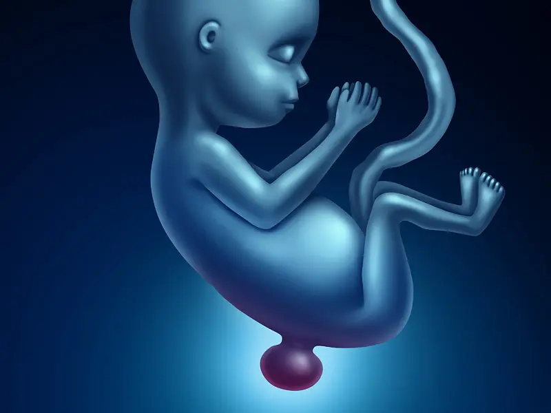
1-Kidney Absence:It occurs in 1 in 1000-2000 births and is usually unilateral. It is generally asymptomatic and is detected incidentally. It is thought to have autosomal dominant (non-sex-linked) inheritance. If it is detected in one family member, other family members should also be evaluated with ultrasonography.
CONGENITAL KIDNEY ANOMALIES
1-Kidney Absence:It occurs in 1 in 1000-2000 births and is usually unilateral. It is generally asymptomatic and is detected incidentally. It is thought to have autosomal dominant (non-sex-linked) inheritance. If it is detected in one family member, other family members should also be evaluated with ultrasonography. Theoretically, the increased filtering function in the absent kidney to compensate for the function of the missing kidney could cause progressive damage to the existing kidney, leading to hypertension; however, kidney function remains normal in affected individuals for many years. Although mild proteinuria and high blood pressure may develop in affected individuals, the clinical significance of this is still unknown. Unilateral congenital absence of the kidney is associated with other anomalies of the kidney and urinary system. Vesicoureteral reflux (VUR, leakage of urine from the bladder to the kidney) is the most common anomaly in one-third of these patients, along with other urinary system anomalies. Anomalies in extrarenal organs are also seen in another one-third of patients. In congenital monocytic individuals, high blood pressure is observed in 16% of cases, and microalbumin (protein) excretion in urine is seen in 21%. The glomerular filtration rate (GFR, the kidney's filtering speed) is below 60.
2- Renal Dysplasia and Multicystic Kidney:In newborns, one kidney appears irregular due to numerous cysts. The urinary tract (ureter) is usually absent or underdeveloped. Often, the other kidney is also affected. It is most frequently accompanied by narrowing at the outlet of the other kidney. In 60% of affected kidneys, there is also an obstruction in the urinary tract. It can be detected in the early stages of pregnancy.
3- Potter Syndrome:Oligohydramnios occurs in 1 in 4000 births, with irregular distributions or as an autosomal recessive (non-sex-linked) genetic inheritance. In pregnancy, oligohydramnios is characterized by a decrease in amniotic fluid, the fluid surrounding the baby. This fluid deficiency is caused by the absence of both kidneys. However, oligohydramnios can also occur in newborns with polycystic kidney disease, small kidney size, or obstructive urinary tract diseases. Due to oligohydramnios, the lungs cannot develop, and the baby has a typical facial appearance. Survival is impossible with the absence of both kidneys. The absence of both kidneys can be detected prenatally with ultrasonography.
4- Underdeveloped Kidney (Hypoplasia):One kidney may be small, or both may be small. Low-grade growth deficiency is common and usually asymptomatic. The kidney simply appears smaller than normal. Because the blood vessels in small kidneys are also small, they can cause hypertension that may require kidney removal. In cases of underdevelopment affecting only one part of the kidney (also called Ask-Upmark kidney), severe hypotension is seen at a young age.
5- Multiple Kidneys:An extra kidney is extremely rare and should not be confused with a condition called duplication, where the urinary tract from the kidney is double instead of single. It is usually detected by chance. Fewer than 100 cases have been reported to date. It is more common on the left side. Anomalies in the urinary tract connected to the extra kidney and other genital anomalies are frequently associated. Swelling in the urinary tract (hydronephrosis) is common in cases of extra kidneys.
6- Polycystic Kidney Disease:Adult-onset polycystic kidney disease (APD) occurs in approximately 1 in 400-1000 births. In 10% of patients, it leads to kidney failure requiring dialysis or kidney transplantation. It generally affects adults, and these patients may also have cysts in the liver and pancreas. Chronic hypertension, intracranial aneurysms, and heart valve abnormalities, particularly mitral valve prolapse, are common. Kidney function deteriorates progressively with age.
7- Infantile type polycystic kidney disease:Infantile polycystic kidney disease (ACD) occurs in 1 in 20,000-40,000 births and is genetically inherited via an autosomal recessive pattern. Affected fetuses are born with oligohydramnios and their faces appear similar to those in Potter's disease; some are diagnosed with respiratory failure. 40% of patients have severe liver and kidney failure, and biliary tract problems are seen in 30% of patients. This condition affects both kidneys.
8- Horseshoe Kidney (Adhesion kidney anomalies):Most kidneys are fused at their lower ends. It is twice as common in males, and the renal outlet is usually narrowed. Most fused kidneys are located lower than they should be. It occurs in 1 out of every 600 births. These patients are prone to kidney stones, renal outlet obstruction, infection, and tumor development. Children present with urinary tract infections, abdominal masses, or blood in the urine.
9-Ectopic Kidney (Kidney in an abnormal location):In simple ectopia, the kidney is located lower than normal. The urinary tract from the affected kidney is usually tortuous. In 30% of patients, the renal outlet is narrowed. These kidneys are prone to infection, stones, and urinary tract obstruction. The incidence is 1 in 2200-3000. Non-functional ectopic kidneys may need to be removed. A rarer positional anomaly than simple ectopia is when one kidney is attached to the other without fusion (cross ectopia).
10- Medullary Sponge Kidney:It occurs in 1 in 5000-20000 births. Cystic dilatations are present in the collecting systems of one or both kidneys. Nephrocalcinosis and recurrent stone disease are common. There are also forms inherited with an autosomal dominant trait. It is frequently associated with other anomalies of the urinary tract.
11- Trisomies (Extra Chromosomes) and Congenital Anomalies of the Kidney: TrNot all isomyomas cause kidney anomalies, but hydronephrosis (dilation of the urinary ducts in the kidney), horseshoe kidney, double collecting system, kidney cysts, and/or cystic dysplasia or hypoplasia (congenitally small kidney) are common in cases of excess chromosomes 13, 18, and 21.
ANOMALIES OF THE URETER (THE LONG URINARY TRACT BETWEEN THE KIDNEY AND THE BLADDER) AND THE URETEROPELVIC JUNCTION (THE POINT WHERE THE URETER LEAVES THE KIDNEY).
1- Ureter atresia (underdeveloped ureter):The ureter is either absent or does not reach the bladder, ending in a blind end. It is a rare condition where the kidney on the same side is absent and the cyst is multicystic. The affected kidney is often non-functional. Diagnosis can be made with prenatal ultrasonography. Bilateral cysts are incompatible with life. Unilateral cysts often do not cause symptoms but can lead to hypertension.
2-Double Ureter (ureter duplication):Occurring in approximately 1% of cases, it is the most common congenital anomaly of the urogenital system. The double ureters may continue to the bladder or may merge before reaching it. In cases where the ureters do not reach the bladder, vesicoureteral reflux or UPJ stenosis is common. In cases with double ureters extending to the bladder, ectopic ureter (the ureter opening to an opening outside the bladder) and ureterocele (a sac-like swelling at the bladder end of the ureter) are seen. Unlike other anomalies, it is six times more common in girls. It is often bilateral. Persistent urinary tract infections and subsequent chronic kidney failure may develop. It increases the risk of kidney infection 20-fold in children, while in adults, it is often asymptomatic. It is inherited in an autosomal dominant manner.
3-Ureterocele:It is a sac-like dilation in the bladder or at the end of an ectopic ureter. It is more common in girls than boys. It is bilateral in 10% of cases. It may be asymptomatic or may occur in conjunction with vesicoureteral reflux and/or bladder outlet obstruction. It is rare.
4-Ectopic Ureter (where the end of the ureter opens into an opening outside the bladder):It is often associated with ureterocele and ureteral duplication. In male children, if the ureter opens into the sperm ducts, it may be seen with testicular infection. In female children, if the ureter opens into the urethra, continuous dribbling of urine occurs.
5- UPJ Stenosis:This is an obstruction at the point where the renal outlet joins the ureter. It occurs in 1 in 000-1500 births and is the most common cause of renal swelling in the womb. It can develop due to many reasons, including abnormalities in the ureteral junction, thickening of the ureteral muscle layer, fibrous tissue formation, and narrowing at the ureter's entry point into the kidney. Prolonged persistence can lead to kidney infection (pyelonephritis), hydronephrosis, and chronic kidney disease. Diagnosis can be made through maternal blood tests. Resistance to the passage of urine from the kidney to the ureter causes dilation of the intrarenal urinary tract and impaired kidney function. 80% of prenatally diagnosed hydronephrosis cases are due to UPJ stenosis. Pain and vomiting are seen in children. It often requires surgery, and sometimes it may not show symptoms until adulthood. It is twice as common in boys and twice as common in the left kidney compared to the right. In patients with kidney stones, UPJ stenosis can also develop later due to trauma or external compression of the UPJ.
6- Prune Belly Syndrome:It occurs in 1 in 100,000 live male births. The classic three findings are: urinary system anomalies, underdeveloped abdominal muscles, and bilateral undescended testicles. Oligohydramnios (low amniotic fluid), bilateral hydronephrosis, and kidney developmental anomalies are present. The bladder is noticeably distended, and its muscle layer is insufficiently developed. Due to this distended bladder, the testicles cannot descend into the scrotum, and the intestines are not in their normal position. The distension disappears after emptying, and the anterior abdominal wall appears wrinkled like a prune due to the underdeveloped abdominal muscles. It gets its name from this appearance. It can be associated with a wide range of heart, lung, and orthopedic problems, including foot anomalies, underdeveloped lungs, Potter's face, and imperforate anus. The cause is unknown. The bladder can be emptied in the unborn baby, thus preventing conditions related to oligohydramnios; however, stillbirth or death weeks after live birth is common. 96% of patients are male. Reaching adulthood after anterior abdominal wall and urinary system surgeries is rare.
8- Megaureter caused by obstruction:It occurs due to an obstruction at the point where the ureter enters the bladder. It is four times more common in males than females and three times more common on the left side than the right. It is often associated with absence or abnormal development of the contralateral kidney. It can be detected by ultrasound in utero. It can manifest with blood in the urine, abdominal pain, and infection.
BLADDER ANOMALIES
1- Bladder exstrophy:The absence of the anterior bladder wall, resulting in the ureter opening into the anterior abdominal wall, is a common anomaly, especially when accompanied by other abnormalities such as epispadias. Surgical repair is required. Bladder exstrophy and epispadias are variations in midline abdominal developmental deficiencies. Sometimes, hereditary forms are observed.
2. Failure of the urachus to close:It is a congenital anomaly characterized by a cavity just behind the navel that can cause infection.
3- Bladder shrinkage:VUR is a congenital anomaly characterized by balloon-like structures that protrude outwards due to a missing area in the bladder wall, along with symptoms of bladder irritation.
ANOMALIES OF THE PENIS AND URETHRA (URINARY TRACT) IN MEN
1- Absence of a penis (aphalia):It is a very rare anomaly with urological and psychological consequences. In 50% of patients, other urogenital anomalies such as undescended testicles, kidney absence, and kidney maldevelopment are also observed. Gender determination should be performed after surgical correction of life-threatening anomalies.
2. Penis size (megalophallus):It is the rapid growth of the penis during childhood due to high production of male hormones (testosterone).
3- Small penis size (Micropenis):Aside from being small, there is no deformity in the shape of the penis; it is defined as the length of the penis, when extended, being more than 2.5 standard deviations smaller than the average. It should not be confused with buried penis, where the penis length is normal. The most common causes are: Hypogonadotropic hypogonadism (failure of the pituitary/hypothalamus to release hormones that stimulate the testes): Seen in hypothalamic disorders such as Kalmann syndrome and Prader-Willi syndrome. Hypergonadotropic hypogonadism: The testes do not convulse due to reasons such as gonadal abnormalities. Micropenis of unknown cause and hormonal disorders cannot be identified.
4- Urethral stricture (narrowing of the part of the urethra inside the penis):It is uncommon. In severe cases, damage develops due to increased pressure on the bladder and kidneys.
5- Posterior urethral valve (PUV) on voiding cystographyThis is the most common cause of lower urinary tract obstruction in male newborns and fetuses. Diagnosis is usually made prenatally. It consists of a convolution in the cells lining the urethra at the beginning of the portion of the urethra that passes through the prostate. These thin membrane structures open during urination, causing obstruction. Urine flow is slow, drip-like, and urinary tract infections are frequent. It can cause significant kidney damage and kidney failure in infancy. The decision for surgery is made based on prenatal diagnosis, amniotic fluid volume, gestational age, and kidney function.
6- HypospadiasHypospadias is a condition where the urethra is located lower than where it should be at the tip of the penis. In 90% of cases, the urethral opening is at the very tip of the penis, close to its normal location. In 10% of cases, the urethral opening is located far from the tip. In most cases, the lower part of the foreskin is underdeveloped. Downward penile curvature is present in 10% of cases where the opening is near the tip and in 50% of cases where it is far from the tip. Other congenital urogenital anomalies, most commonly undescended testicles (10%), are also seen. Therefore, hypospadias is considered a disease of sexual differentiation. It occurs in 18 out of every 10,000 live births of males worldwide. Its frequency increases with very low birth weight. Older children often complain of urine splashing on them while urinating. Inguinal hernias are also present in 9-15% of cases. Hypospadias is also present in other family members in 7% of cases. Hypospadias is named according to the location of the urethral opening; the closer it is to the end, the easier the treatment.
7-Epispadias:This is a condition where the urethra terminates at a point on the penis due to a lack of formation on the side facing the body. Unlike hypospadias, it is a completely different embryological developmental disorder. It can also occur in female infants. It can be near the tip of the penis or severe enough to cause bladder exstrophy. It occurs in 1 in 120,000 births in males and 1 in 450,000 births in females. In male infants, it causes posterior curvature of the penis. Urinary incontinence is common. Surgery is performed to reposition the urethral opening and restore the normal appearance of the external genitalia.
TESTICULAR ANOMALIES
1- Hypogonadism (low testicular function):The testicles are small, and there is no sexual appearance, sexual drive, or erection. Patients are tall, with long arms and legs. It may be related to undescended testicles and Klinefelter syndrome, a chromosomal disorder. Patients may also have intellectual disability and other comorbidities.
2- Ectopic testis (testis in an abnormal position): TThis condition develops when the testicle does not follow its normal path before descending into the scrotum; it is different from undescended testicles. It is rare, and both testicles may be found on the same side. Possible locations include the lower abdomen, upper thighs, and the area under the skin of the penis. It is often associated with an indirect inguinal hernia. The testicle may be ectopic, while the epididymis, another organ inside the scrotum, may be located in the scrotum.
3- Undescended testicles:The descent of the testis into the scrotum is interrupted. This is the most common congenital anomaly in newborn males. It occurs in 2-4% of males, and its frequency increases in premature births. The exact cause is unknown. It can be classified as palpable or non-palpable on examination. Intervention is considered depending on the location of the testis. If the testis can descend into the scrotum on examination, it is monitored. If the testis is not palpable on bilateral examination, problems with sexual differentiation such as hypospadias are investigated, and hormonal system and genetic evaluation may be necessary. The surgical procedure of bringing the undescended testis into the scrotum is called orchiopexy, and the most suitable time is between 6-12 months of age. After this period, varying degrees of damage occur in the sperm-producing cells in the testis. Even if performed in time, the risk of fertility problems after orchiopexy is 33% in unilateral orchiopexy and 66% in bilateral orchiopexy. The risk of testicular tumors is also 5-10 times higher than normal.
4- Noonan syndrome:It is a congenital disease that occurs in 1 in 1000-2500 live births and is characterized by heart disease, short stature, growth retardation, undescended testicles in males, bleeding predisposition, and a typical facial appearance.
5- Spermatosel: Spermatocele is a common congenital anomaly. It is a painless cystic mass palpable in the posterior part of the testicle. It is usually smaller than 1 cm and generally does not require treatment.
6- Varicocele:Varicocele is the enlargement of the vascular structures surrounding the testicle called the pampiniform plexus. It develops slowly, can be present at birth, is rare in children under 10, generally begins with puberty, and is seen in 14-20% of adults. Although not always the case, it may be caused by the congenital absence of the valves that should be present in the left testicular veins. In most cases (78-93%), it is on the left side; if it is unilateral on the right, it should be considered in conjunction with major vessel diseases. Fertility problems are seen in 20% of adults with varicocele. Conditions for which surgery is recommended include: testicular shrinkage along with varicocele, presence of testicular disease other than varicocele affecting fertility, presence of impaired sperm quality, bilateral varicocele, and painful varicocele.
7- Hydroseal:Hydroceles are an increase in the fluid surrounding the testicles in the scrotum (sac). At birth, the scrotum is swollen, either unilaterally or bilaterally. The size of the swelling varies. It is often not tense and appears bluish in color. It usually resolves within 24 months; hydroceles detected after this period should be surgically treated. If an inguinal hernia is also present, surgery is recommended before the 24-month mark. Hydroceles can be a common congenital anomaly, but they can also develop later due to trauma or other causes.
GONADAL DYSGENESIS:
Turner syndrome is a sexual development disorder resulting from anomalies in sex chromosomes affecting the testes in males or the ovaries in females, or mutations in genes that influence the appearance of these organs. It can manifest in a wide range of forms, from a normally masculine-looking male to an intersex or female-looking child. Gonadal tumors (testes and ovaries) are common in these children. Gonads with developmental abnormalities can cause a wide range of intersex appearances with varying degrees of maturation or abnormality. Patients carrying the Y chromosome have an increased risk of developing germ cell tumors called gonadoblastoma. In Turner syndrome, which can be seen with mosaic chromosome numbers such as 45X or 46XY, surgical removal of the gonads is recommended to prevent tumor development. Kidney anomalies are frequently observed. Gonadal abnormalities may be unilateral, with the other gonad being normal.
Turner Syndrome:It occurs in 1 out of 2500 live female births, and is frequently associated with growth retardation, abnormal gonadal development, heart anomalies, kidney anomalies, and autoimmune diseases. Urinary infections and hypertension are also more common in patients. Most abnormalities in Turner syndrome are caused by the absence of a specific genetic region on the X chromosome.
Swyer Syndrome:46XY is a gonadal developmental disorder that causes male appearance despite a female chromosomal appearance. It occurs in 1 in 20,000 to 80,000 births. In 30% of patients, it causes germ cell tumors in the form of gonadoblastomas in the ovaries, resembling streaks. In adulthood, the girl does not menstruate, and the uterus is small despite appearing normal.
SEXUAL DEVELOPMENT DISORDERS
This is the new name for disorders formerly classified as intersex disorders. The name was changed after the molecular genetic causes of abnormal sexual development became known. There are still disagreements regarding clinical approaches. In infants born with intersex external genitalia, the most urgent action is to determine gender. External genitalia can include intersex organs, female genitalia with clitoral hypertrophy, male external genitalia with undescended testicles, micropenis, or hypospadias. Causes include complete or partial androgen insensitivity, 5-alpha reductase enzyme deficiency, congenital adrenal hypofunction, developmental disorders involving both ovaries and testes (true intersex), and other causes. By determining a gender for the patient, personality and sexual development are guided accordingly.
Situations where a sexual development disorder is suspected include:
- In a distinctly masculine appearance:Severe hypospadias with a bifurcated scrotum; undescended testicles on one or both sides with hypospadias; absence of both testicles on examination in a seemingly normal newborn.
- In a distinctly feminine appearance:The presence of gonads that are not palpable on examination, clitoral enlargement, and abnormalities in vaginal development.
- Situations where outward appearance cannot be clearly distinguished:Intersex external genitalia, and in female offspring born with intersex external genitalia and chromosome 46XX, adrenal gland disorders leading to salt wasting require urgent attention.
The information here is for general education only and is not medical advice. Please consult your physician for personalized treatment.