









Urethral Stricture
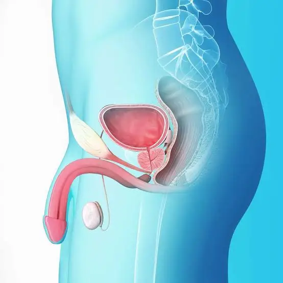
The urethra is the urinary canal between the tip of the penis and the bladder that allows urine to be carried out of the body. Urinary tract stricture (urethral stricture) is a mechanical disease that prevents urine from comfortably exiting the bladder and being discharged outside, ultimately causing urine to accumulate in the bladder and preventing the bladder from fully emptying.
What Is Urinary Tract Stricture, Urethral Stricture?
The urethra is the urinary canal between the tip of the penis and the bladder that allows urine to be carried out of the body. Urinary tract stricture (urethral stricture) is a mechanical disease that prevents urine from comfortably exiting the bladder and being discharged outside, ultimately causing urine to accumulate in the bladder and preventing the bladder from fully emptying.
Urethral stricture is a disease affecting approximately 0.5–1% of men and results from damage to the urinary tract cells (urethral epithelium) for various reasons, leading to fibrosis (scar formation) in the epithelium or in the structure forming the body of the urinary canal. The stricture occurs due to injury or damage to the urethra and surrounding tissue. For example, just as stretching and thickening occur in injured areas of the skin, similarly scar tissue forms in urethral injuries. When this stretching and thickening block the narrow lumen of the urethra, urine cannot pass through the urethra or passes with difficulty depending on the degree of narrowing, and therefore urine accumulates in the bladder and does not empty completely.
Trauma, certain medical applications such as radiotherapy for prostate cancer, some surgical operations performed in this area, and certain infections such as gonorrhea may cause urethral stricture. The causes of urethral stricture vary according to the patient’s age and the location of the stricture. Anterior urethral strictures mostly occur due to infection (40%), medical intervention (iatrogenic-40%), or trauma, whereas posterior urethral strictures occur iatrogenically after pelvic fractures or surgical interventions. Sometimes no cause can be found (15–20%). Strictures may range in length from a few millimeters to several centimeters between the bladder and the tip of the penis.
What Are the Symptoms of Urinary Tract Stricture?
Difficulty starting urination
Slow or weak urine flow
Dribbling after urination
Pain or burning during urination
Blood in the urine
Increased frequency of urination
Inability to completely empty the bladder
Urinary tract infections
In severe cases, the urethra may become completely blocked and lead to acute urinary retention requiring emergency medical intervention. Sexual dysfunctions such as difficulty achieving or maintaining an erection or painful ejaculation may also be seen due to urethral stricture. Sometimes patients may present with recurrent urinary tract infections, prostatitis, epididymitis-orchitis, or bladder stones.
How Is Urinary Tract Stricture Diagnosed?
Clinical history
Symptom scoring
Physical examination
Uroflowmetry and ultrasonography, residual urine amount
Urethrography
Urethrocystoscopy (in some cases)
Before treatment of urethral stricture, a detailed history regarding the cause should be obtained. In physical examination, evaluation of urinary meatus pathologies and palpation of scar tissues in the anterior urethra in cases called lichen sclerosus are important. In patients with urethral stricture, maximum urine flow rate and urination pattern should definitely be evaluated with a urination test (uroflowmetry). Ultrasonography may be necessary to evaluate bladder pathologies and measure the amount of residual urine that cannot be emptied. Retrograde urethrography (RUG) may be performed to evaluate the exact location and length of the urethral stricture. Retrograde urethrography is the gold standard especially for diagnosing anterior urethral strictures. However, since urethrography is insufficient in posterior urethral strictures and bladder neck pathologies, a combination of voiding cystourethrography and urethrography should be performed in these cases.
In cases where diagnosis cannot be clarified, urethrocystoscopy may be necessary to clarify the stricture. Flexible cystourethroscopy performed simultaneously through the external urinary opening or cystostomy route may be an important diagnostic tool in measuring the location and length of the stricture. After the location and length of the stricture are clarified, the method and timing of intervention are planned.
How Is Urinary Tract Stricture, Urethral Stricture Treated?
Treatment in urethral strictures may vary according to the cause, location, number, length of the narrowed area, severity-density of the stricture (degree of spongiofibrosis), previous treatment attempts, and the patient’s age. Relatively short simple strictures are treated endoscopically, whereas long-complex strictures are treated with one-stage or two-stage open surgery (urethroplasty).
1- Urinary Tract Dilatation
It can be performed with metal dilators, balloon dilatation, or catheterization. This treatment method may provide temporary relief to the patient by opening the scar formed in short segment strictures (<1 cm). It is generally applied to patients with additional diseases who cannot tolerate another intervention and who have limited life expectancy. However, in short strictures without severe scar tissue, it may rarely provide complete treatment.
2- Drug-Coated Balloon Treatment in Urinary Tract and Urethral Strictures, Optilume Treatment
Optilume (drug-coated balloon catheter) is a minimally invasive drug-coated balloon application used in the treatment of urinary tract strictures (urethral strictures). Optilume impregnated with a drug called paclitaxel is a drug-coated balloon catheter or stent that expands the urinary tract with its balloon and directly delivers the impregnated drug to the stricture. It is a relatively new procedure that significantly reduces the possibility of recurrence of urinary tract stricture, promising results are being obtained, and it is becoming an increasingly popular option for patients with urethral stricture.
With this balloon catheter impregnated with the drug called paclitaxel, in addition to expanding urinary tract strictures, the loaded drug is directly delivered to the stricture and thus significantly reduces the possibility of recurrence of the stricture. Recovery after the procedure is rapid and the procedure is relatively painless. Most patients can return to normal activities within a few days after the procedure. In the procedure, a small flexible balloon catheter or stent placed into the urinary canal first expands the stricture with the help of the catheter balloon, and meanwhile the drug is released into this stricture area.
One of the most important advantages of Optilume is that it is a minimally invasive procedure, which means less damage to the tissue surrounding the stricture. Optilume also has the advantage of a faster recovery process and causing less pain and discomfort for the patient. In addition, since the procedure is minimally invasive, the risks of complications such as infection, bleeding, or damage to surrounding tissue are also reduced. It is generally an outpatient procedure. Thanks to the drug it contains, it prevents poor healing that causes re-narrowing such as scar tissue.
3- Internal Urethrotomy, Closed Endoscopic Surgery
Closed Endoscopic Surgery is the procedure of cutting the short urethral stricture with a cold knife using an endoscope called a urethroscope. This method has been widely used for approximately fifty years. Various lasers other than this cold knife can also be used. In general, the success of laser urethrotomy is reported to be the same as with the cold knife.
After Closed Endoscopic Surgery, the scarred urethral tissue is left to wound healing and urinary tract continuity is re-established. If normal healing occurs before scar tissue formation, the procedure is successful; otherwise, if wound healing occurs more rapidly, recurrence of the stricture is almost inevitable. With careful patient selection (soft short bulbar stricture <1 cm), approximately 80% success can be achieved, however in strictures longer than 2 cm, multiple, dense-severe strictures, and strictures close to the external opening, it generally results in failure. Open surgery (urethroplasty) should be preferred in cases recurring within 3 months after internal urethrotomy, recurring more than three times a year, or recurring after two failed interventions.
Long-term clean intermittent self-catheterization by the patient may be required after Closed Endoscopic Surgery. Placement of a permanent catheter should be recommended in patients with other serious diseases, limited life expectancy, or those who cannot undergo another surgical procedure. Although the success of Closed Endoscopic Surgery varies, a success rate between 70–80% has been reported. Despite varying success rates, due to its easy applicability, low cost, short hospital stay, and repeatability, internal urethrotomy is preferred as the most frequently used method.
Complications
The most common main complication in internal urethrotomy surgery is recurrence of the stricture. Other complications (0.5–5%) generally include bleeding, hematoma, and epididymo-orchitis (testicular inflammation). In some rare cases, deep incisions may cause fistula formation leading to erectile dysfunction.
4- Urethroplasty
Urethroplasty is the most effective method for the complete treatment of urethral strictures and is considered the gold standard. In this method, after removal of the stricture area, urethroplasty is performed either by end-to-end suturing or by using grafts taken from other tissues of the body (oral mucosa, graft taken from leg vessels).
Removal of the Stricture and End-to-End Anastomosis (Excision and Primary Anastomosis)
The aim of this treatment method is to completely remove the scar tissue (fibrotic tissue) and then appropriately reconnect the urethra end-to-end. With this technique, approximately 90–95% success is achieved in bulbar urethral strictures below 2 cm. In strictures longer than 2 cm, it may be necessary to carefully separate the two corpus cavernosum tissues in order to reduce end-to-end anastomosis tension.
Augmentation Urethroplasty
This treatment method is generally preferred in cases longer than 2 cm where end-to-end suturing is not possible and urethroplasty is not suitable. This method is performed in one or two stages. In augmentation urethroplasty, recurrence of stricture has been reported at a rate of 14–15%. The use of grafts in urethroplasty shows no difference in terms of recurrence of stricture. Penile skin, scrotal skin, oral mucosa, bladder mucosa, and colonic mucosa may be used for grafting. Among these, oral mucosa is the most preferred material due to ease of harvesting, absence of hair-bearing skin, low complications, and high success rates. Oral mucosa may be obtained from the cheeks, lips, or tongue. Reported complications related to oral mucosa graft harvesting include bleeding, pain, infection, swelling, and salivary duct injury. Some patients may experience temporary difficulty opening their mouth.
Two-Stage Urethroplasties
These are applied in penile urethra and especially in cases with failed hypospadias surgery or lichen sclerosus. After the first stage, severe stricture due to graft scarring may be seen at a rate of 10–39%. Therefore, it is necessary to wait 3 to 6 months for the second stage.
The information here is for general education only and is not medical advice. Please consult your physician for personalized treatment.