









URS
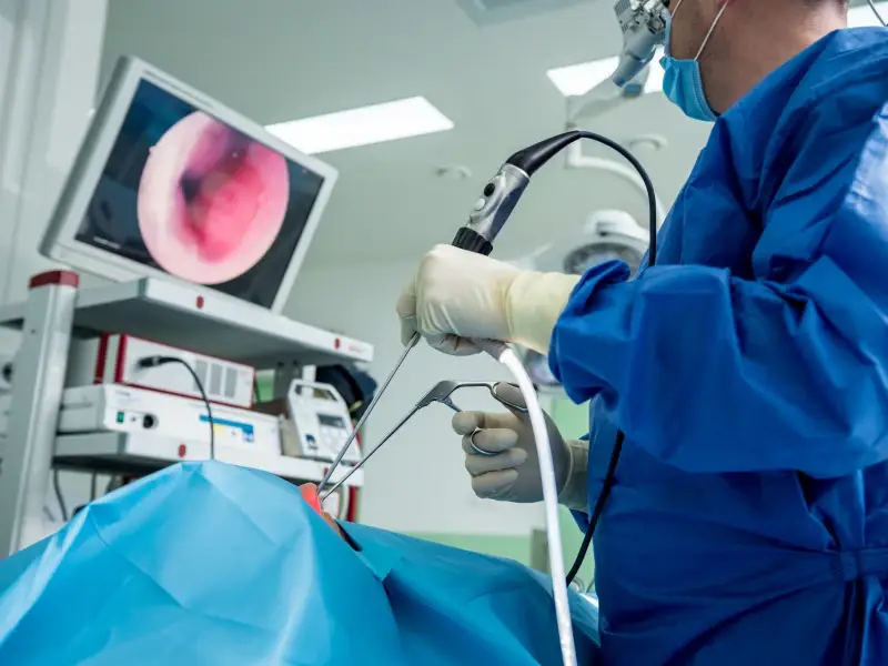
Among the surgical options for patients with kidney stones are extracorporeal shock wave lithotripsy (ESWL), closed endoscopic ureteroscopy (Flexible URS), and percutaneous nephrolithotomy (PNL), which is performed through a needle inserted from the back. The condition of the kidney, the type, location, and size of the stone, as well as the physician’s preference, play an important role in determining the surgical approach.
Laser Kidney Stone Surgery, Closed Kidney Stone Surgery, Laser Kidney Stone Fragmentation, Endoscopic Kidney Stone Surgery, What is Flexible URS? How Is It Performed?
Among the surgical options for patients with kidney stones are extracorporeal shock wave lithotripsy (ESWL), closed endoscopic ureteroscopy (Flexible URS), and percutaneous nephrolithotomy (PNL), which is performed through a needle inserted from the back. The condition of the kidney, the type, location, and size of the stone, as well as the physician’s preference, play an important role in determining the surgical approach.
Ureteroscopy, also known as ureterorenoscopy (URS), is a commonly used endoscopic and camera-assisted diagnostic and treatment method for diseases located in the kidneys and urinary tract (ureters). Due to developments in ureteroscope size and flexibility, video imaging, miniature baskets and instruments, and the emergence of holmium laser stone fragmentation, the closed endoscopic treatment of kidney stones has undergone significant evolution. More than 25% of all kidney stone surgeries are now performed using small flexible URS, namely ureteroscope technology.
What Should Be Done Before Laser Kidney Stone Surgery?
During the first clinical examination before surgery, all imaging films and reports such as CT scans, intravenous pyelography (IVP), sonograms, or MRI are obtained and carefully reviewed by the surgical team. If necessary, blood and urine tests are performed, medical history is reviewed, and a physical examination may be conducted. The patient is then evaluated for ureteroscopy, namely laser kidney stone fragmentation. After this decision, surgical preparation begins. An anesthesia consultation is performed before surgery. For anesthesia preparation, the anesthesia team will conduct a physical examination, ECG, blood count, coagulation tests (INR, PT, PTT), biochemical blood tests, and urinalysis.
Closed kidney stone surgery or ureteroscopy is the only minimally invasive stone surgery that can even be performed while the patient is taking active blood-thinning medications. However, even in this case, if medically possible, discontinuation of all blood-thinning medications before surgery is generally preferred.
How Is Laser Kidney Stone Surgery and Laser Kidney Stone Fragmentation Performed?
After the patient is asleep under general anesthesia, a thin illuminated camera-assisted tube (flexible ureteroscope) is passed through the urinary canal and bladder into the ureter, reaching the location of the stone. If the stone is small, it may be grasped with a basket device and removed intact through the ureter. If the stone is large and/or the ureter diameter is narrow, the stone needs to be fragmented, usually using a holmium laser. After the stone is broken into small fragments, these fragments are generally removed using instruments called baskets or forceps. In most cases, a temporary ureteral stent is placed to ensure proper drainage of urine from the kidney after surgery and to reduce the damage caused by the stone and the procedure. This stent can easily be removed a few weeks later.
For stones located inside the kidney, laser kidney stone fragmentation may also be performed using flexible ureteroscopy, also known as ureterorenoscopy. Similar to ureteral stones, kidney stones are fragmented with a holmium laser and may be removed using basket instruments. Sometimes the kidney stone is fragmented into very small pieces (sand-like particles) that cannot be collected with a basket. Again, a temporary ureteral stent is usually placed to ensure proper drainage of urine after surgery and to reduce the damage caused by the stone and the procedure. This stent can easily be removed a few weeks later.
Finally, if the ureter is too narrow for the ureteroscope to pass through, the procedure is generally ended by placing only a stent. The stent allows the urinary tract to widen within a few weeks and become suitable for the procedure. Afterwards, the stone surgery is performed as described above and completed. Ureteroscopy, namely laser kidney stone surgery, is generally performed as an outpatient procedure. However, some patients may need to stay in the hospital overnight if the procedure is prolonged or difficult.
You may watch an animation of Laser Kidney Stone Surgery by clicking this link:
https://www.youtube.com/watch?v=9X7Arktn96E&t=24s
What Are the Possible Complications During or After Laser Kidney Stone Surgery?
As with any major surgery, although rare, complications may occur during or after laser kidney stone surgery. Potential risks and complications related to this operation include:
Stent pain: Approximately 50% of patients who undergo ureteroscopy and receive a stent experience “stent pain,” which is by far the most common complaint after ureteroscopy. The stent is a soft plastic tube that allows the kidney to drain into the bladder independently of edema or obstruction. The stent may irritate the bladder lining, causing urgency and overactive bladder symptoms. It may also cause reflux of urine from the bladder back to the kidney during urination, resulting in warmth, tingling, and severe pain in the operated kidney area. These complaints generally disappear after the stent is removed a few weeks later.
Residual stone fragments: Depending on the original stone size and location, residual stones may remain in the kidney or ureter in up to 40% of patients after laser kidney stone surgery. These fragments may pass spontaneously over time or may be monitored during follow-up visits.
Ureter injury: Injury to the ureter is the most common complication during ureteroscopy surgery. The risk of complete perforation is approximately 1 in 1000, while the rate of partial injury is about 1.6%. Nearly all cases heal with prolonged stent placement (2–4 weeks). In cases of major injury, the procedure is stopped and laser stone surgery is repeated after sufficient healing time.
Ureteral stricture and avulsion: Ureteral strictures and ureteral avulsion (complete separation of the ureter from the kidney) are among the most feared complications of endoscopic kidney stone surgery. Fortunately, due to the development of thin-caliber ureteroscopes and increased surgical experience, the risks of avulsion (0.05%, 1/2000) and stricture (0.2%, 1/500) are rare.
Bleeding and infection: Bleeding and infection may occur in approximately 5% of patients after laser kidney stone surgery. However, most cases are self-limiting and improve with hydration and antibiotics.
What Is the Recovery Process After Laser Kidney Stone Surgery?
Immediate postoperative period: After surgery, the patient is first taken to the recovery room. If a urinary catheter (Foley catheter) was placed during surgery, it may be removed once the patient is fully awake and mobile. The patient is then transferred to the regular room, pain is controlled if present, and after the patient urinates for the first time, discharge may be considered. With time and fluid intake, the urine color should gradually change from watermelon red to pink and eventually clear. Due to the instrumentation used during the procedure, most patients are prescribed oral antibiotics for 4–5 days to prevent urinary tract infection.
Postoperative pain: After endoscopic kidney stone surgery, most patients may feel mild to moderate pain in the flank and/or bladder area. This is generally well controlled with oral pain medications.
Ureteral stent (Double J stent): After ureteroscopy, a small tube called a ureteral stent is almost always placed. This stent facilitates the passage of urine from the kidney to the bladder. The stent is generally removed easily after 2–3 weeks.
Nausea: Nausea is quite common after any surgery involving general anesthesia. It is generally temporary and self-limiting.
Showering: Patients may shower immediately after discharge from the hospital.
Activity: Patients may resume driving once they stop taking pain medications. Most patients can return to normal daily activities within 5–7 days after the procedure. However, many patients report increased fatigue and discomfort due to the ureteral stent, which may limit some activities.
Diet: Most patients are advised to consume mainly liquid foods during the first 24 hours after laser kidney stone surgery because bowel function may slow down due to the surgery and general anesthesia. Afterwards, patients may gradually return to a normal diet as tolerated.
Fatigue: Fatigue is very common after surgery and usually resolves within a few days.
Constipation/Gas cramps: Due to anesthesia, bowel problems may occur for several days after laser kidney stone surgery. Suppositories and stool softeners may help relieve this issue. Pain medications may also contribute to constipation, so patients are encouraged to discontinue pain medications as soon as tolerated.
Frequently Asked Questions About Laser Kidney Stone Surgery, Closed Kidney Stone Surgery, Laser Kidney Stone Fragmentation, and Flexible URS
1- What are the advantages of Laser Kidney Stone Surgery compared to other stone treatment methods?
If the size and location of the kidney stones are appropriate, the advantage of flexible ureteroscopy is that it allows access to every part of the kidney. As long as the ureter is large enough to allow the ureteroscope to pass, there is a high chance that the stone can be fragmented and removed in a single surgery.
Compared to extracorporeal shock wave lithotripsy (ESWL), the kidney or ureteral stone can be directly visualized under direct vision through the ureteroscope, allowing simultaneous fragmentation with laser and removal using a basket or forceps. In shock wave lithotripsy, patients are expected to pass the stone fragments themselves, which may cause additional pain or obstruction. In addition, shock wave lithotripsy may not be able to fragment very dense and hard stones, which are known to be resistant to ESWL. Holmium laser ureteroscopy can fragment almost every type of stone as long as the stone is accessible through the endoscope. Furthermore, ureteroscopy also allows treatment of radiolucent stones such as uric acid stones, which cannot be seen on X-rays.
Compared to percutaneous nephrolithotomy (PNL), the endoscope is advanced through natural body openings, meaning no incision is made on the skin and hospital stay is shorter. Some patient groups that cannot be treated with ESWL or PNL (patients using blood thinners, pregnant women, severely obese patients, airline pilots/astronauts, etc.) may be treated safely and effectively with ureteroscopy.
2- Who is not a good candidate for Laser Kidney Stone Surgery? For whom is this surgery not suitable?
Patients with large stones: Since ureteroscopy requires active removal of most or all stone fragments, treating very large stones (>2 cm) may produce too many fragments, making complete removal impractical or impossible.
Patients with a history of urinary tract reconstruction: The anatomy of patients who have undergone ureteral or bladder reconstruction may not allow passage of the ureteroscope.
Patients who cannot tolerate stents: In patients with a history of intolerance to ureteral stents placed during laser kidney stone surgery, alternative stone treatment methods may be preferred.
3- What are the success rates of Laser Kidney Stone Surgery, namely Ureteroscopy (URS)?
Depending on the size, location, and number of stones, success rates range between 50% and 90%. The surgeon can provide success rates specific to the patient’s stone disease.
4- How long will the stent placed after Laser Kidney Stone Surgery remain in place?
The duration the stent remains in the ureter varies. Your doctor will most likely recommend removal 5–10 days after the procedure. Approximately 50% of patients experience flank fullness (usually during urination) and pain due to the stent. These complaints often decrease over time. Since long-term retained ureteral stents may become calcified, encrusted, lead to chronic infections, chronic kidney obstruction, and eventually loss of kidney function, timely removal of the stent within a few weeks is critically important.
5- What is the ureteral stent placed during Laser Kidney Stone Surgery?
The ureter is the natural tube that carries urine from the kidney to the bladder. A ureteral stent is a specially designed hollow tube made of soft plastic material placed inside the ureter. This tube facilitates urine passage until the obstruction resolves. Stent sizes and lengths vary according to patient characteristics.
6- Why is a stent placed?
Placement of a ureteral stent allows urine to pass from the kidney to the bladder even if the ureter is obstructed (stone, edema, external compression, tumors, clots, etc.). The stent helps prevent pain that may occur due to kidney obstruction while supporting proper kidney function. In addition, ureteral stents help the kidney clear bacterial infections associated with obstruction.
The stent placed after ureter or kidney surgery also contributes to the healing of damage caused by the stone or surgical intervention within the ureter. If a stent is not placed after surgery, ureteral strictures may sometimes develop. Since stents support tubular healing, they help prevent this condition.
Sometimes a stent is placed to allow the ureter to widen over a certain period. This is important for passing instruments used during laser kidney stone surgery or for removing stones. This situation occurs when the ureter diameter is too narrow for instrument passage or when ureteral stones narrow the lumen due to edema or inflammation. Stent placement allows passive expansion of the ureter in preparation for future successful interventions.
7- What are the disadvantages of ureteral stent placement?
Approximately 50% of patients experience some form of stent-related side effect. While some patients experience complaints for only a few days, others may continue to have symptoms throughout the entire stent duration.
Hematuria: Stents may cause blood in the urine at various times. Physical activities such as walking or running may cause the stent to move inside the body, resulting in hematuria.
Pain: Pain related to the stent may be felt in the back (lower back), bladder region, groin, penis in men, urethra in women, and sometimes the testicles. Discomfort or pain may become more pronounced after physical activity and urination.
Bladder spasms: The stent may irritate the inner surface of the bladder, causing more frequent urination during the day and night. These symptoms may sometimes improve with medication.
Urinary incontinence: Rarely, the stent may cause urinary leakage due to bladder spasms. This is generally controlled with oral medications or stent removal.
Stent migration: Stents may move from their intended position to other parts of the urinary tract, causing pain or urinary leakage.
Infection: Since stents are foreign bodies, bacteria may adhere to their surfaces. These bacteria may later be released into the urine, causing infection and fever. Although antibiotics may temporarily treat these infections, recurrence may occur because antibiotics cannot fully penetrate bacteria attached to the stent.
Calcification and encrustation: Stents may sometimes be forgotten by patients or their relatives. Over time, they may become coated with urinary salts and minerals and eventually form very large calcified stones. This may lead to chronic obstruction, pain, chronic infections, and even complete loss of kidney function.
8- How is a stent inserted?
The stent is generally placed under general anesthesia, often during another surgical procedure. A telescope called a cystoscope is advanced through the urethra into the bladder. The stent is then advanced into the ureter through the cystoscope using a guidewire, and its position is confirmed with X-ray imaging.
9- How is a stent removed?
Under local anesthesia in the urology clinic, a special flexible telescope (cystoscope) is advanced through the urethra into the bladder. The ureteral stent is grasped with a holding instrument and removed.
10- How does the stent affect daily life?
Most patients can continue normal daily activities while the stent is in place. However, many patients report increased fatigue and discomfort during the day, which may limit activity levels. Additionally, some patients experience bladder spasms requiring more frequent urination, making travel more uncomfortable or difficult. Ureteral stents do not restrict sexual activity, but the side effects described above may reduce comfort and pleasure.
11- What should be done while the stent is in place?
No special care is required. Drinking at least 1.5–2 liters of fluid per day is encouraged to help dilute the urine. If the stent causes constant severe pain, if urinary tract infection symptoms occur (fever, chills, feeling unwell, painful urination), or if the stent falls out, the patient should contact their urologist.
12- What are the alternatives to ureteral stents?
If the obstruction is expected to be temporary, it may be reasonable not to place a ureteral stent. The surgeon decides during the procedure whether the situation is suitable for a “stent-free” approach.
The information here is for general education only and is not medical advice. Please consult your physician for personalized treatment.