









Kidney Pelvis and Ureter Cancers
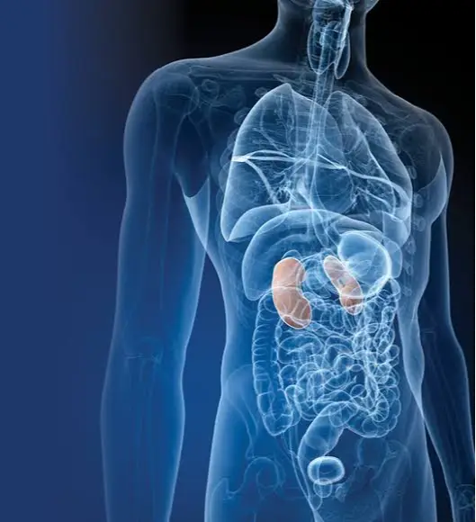
The renal pelvis and ureters form the upper urinary tract and are part of the urinary excretory system.What is the Pelvis?:The renal pelvis is a hollow space in the center of each kidney, also called the renal pool.What is the ureter?:Ureters are thin tubes that connect the kidneys to the bladder.
What are the renal pelvis and ureter, and what is their structure?
The renal pelvis and ureters form the upper urinary tract and are part of the urinary excretory system.What is the Pelvis?:The renal pelvis is a hollow space in the center of each kidney, also called the renal pool.What is the ureter?:Ureters are thin tubes that connect the kidneys to the bladder. Each ureter is approximately 25 to 30 cm long. Urine is your body's liquid waste and is produced by the kidneys. It collects in the renal pelvis and travels down the ureters to the bladder where it is stored. When the bladder is sufficiently full of urine, it is expelled from the body through the urethra (urinary tract).
The walls of both renal pelvises and ureters consist of three distinct layers.
- Mucous membranes:Urothelia (the cells lining the pelvis, ureters, and bladder) can be considered the inner lining of both renal pelvises, ureters, bladder, and urethra (the urinary tract). These urothelial cells, which can stretch and change shape during urine flow, are also called transitional epithelium.
- Lamina propria:The renal pelvis and ureters of both kidneys are located beneath the urothelium, a thin layer of connective tissue containing blood vessels, nerves, and some glands.
- Muscularis propriaThe urinary tract is the thick, outer muscular layer of both renal pelvises and ureters. It consists of involuntary, automatically functioning muscles (smooth muscle). Its function is to propel urine from the kidneys to the bladder.
Other tissues surrounding the kidneys and ureters include:
- Adventitia layer:Loose connective tissue that covers the kidneys and ureters.
- oil layer: A layer of fat surrounding the renal pelvis, kidney, and ureter.
What is Pelvic and Ureter Cancer?
Renal pelvis and ureter cancer, also known as upper urinary tract cancer, are malignant cancers that originate in the cells of the renal pelvis (the renal calyx) and/or ureters (the tubes connecting the kidney to the bladder). These cancers can spread to all layers of the pelvis and ureter, to neighboring tissues, and to other distant parts and organs of the body. Renal pelvis and ureter cancers are 3-4 times more common in men than in women, with an average age of onset of 65.
Renal pelvis, Urothelial Cancers of the Ureters
The renal pelvis, ureters, bladder, and urethra (urinary tract) are lined with cells of the same type called transitional epithelium, and cancers in these areas are collectively called urothelial cancers or transitional epithelial cancers. Due to various reasons, the urothelium cells covering these areas begin to proliferate, grow, and become cancerous, and if left undetected, they continue to spread. Depending on the location, it is called bladder cancer (most common), ureteral cancer if it occurs in the ureters, and renal pelvis cancer if it occurs in the renal pelvis. 90% of cancers in these areas are of this type; the remaining 10% are squamous cell carcinomas and adenocarcinomas, which also originate from the same cells and undergo differentiation. Renal pelvis and ureteral cancer are most common in the renal pelvis, but they can occur simultaneously in several areas of the ureters, or be localized only in the ureters.
Approximately 20% to 40% of patients with urothelial carcinoma in the pelvis and/or ureters also develop bladder cancer, originating from the same cells, either simultaneously or at different times. Therefore, patients diagnosed with pelvic and/or ureteral cancer should be screened for bladder cancer and other cancers in the urinary tract. Conversely, there is a possibility of pelvic and ureteral (upper urinary tract) cancer coexisting or developing at different times in patients with bladder cancer, with a rate of 4%.
Pelvic and/or ureteral cancers (as with bladder cancer) are divided into two groups, superficial (non-invasive) or metastatic (invasive), depending on how far they grow into the walls of these structures.
Superficial (non-invasive) urothelial carcinoma:Superficial urothelial carcinoma refers to cancer that is limited to the inner lining of the pelvis and/or ureter. It can develop as papillary (raised, cauliflower-like) lesions or as flat (stalked) lesions.
- Papillary superficial urothelial carcinomasThey are small, finger-like protrusions and tend to grow into the pelvic and/or ureteral cavity. These superficial papillary urothelial cancers can be low-grade or high-grade. However, small papillary cancers generally tend to be low-grade. These tumors are called papillary urothelial neoplasms with low malignant potential (PUNLMP). Some low-grade tumors also have a small chance of transforming into an invasive, metastatic cancer.
- Flat superficial urothelial carcinomasThese are tumors that grow along the lining of the pelvis and/or ureter. They are usually high-grade and have a higher probability of descending deeper into the layers of the pelvic and/or ureteral wall. This type of squamous urothelial carcinoma more commonly involves carcinoma in situ (CIS) and has a high potential for metastasis.
Disseminated-invasive urothelial carcinoma:Invasive urothelial carcinomas have grown into deeper layers of the renal pelvis and/or ureter wall. They may have extended beyond the wall into the pelvis and/or ureter and grown into adjacent areas.
Sometimes, disseminated-invasive urothelial carcinomas contain a mixture of normal urothelial cancer cells and groups of cells with different characteristics that have been differentiated or altered from these cells, in which case the prognosis of such cancers is worse. These different cell groups may include squamous cells, glandular cells, and small cells.
There are rare subtypes of urothelial carcinoma called variant types. These subtypes generally grow and spread rapidly and tend to have a worse prognosis than normal urothelial carcinoma.
Variant types of renal pelvis and ureter cancer include:
Nested (introverted, layered)
- Microcystic
- Micropapillae
- Lymphoepithelioma-like
- Plasmacytoid
- Sarcomatoid
- Giant cell
- poorly differentiated (underdifferentiated)
- Rich in fat
- Transparent cell
Rare cancers of the renal pelvis and ureter
Squamous cell carcinoma:Squamous cell carcinoma of the pelvis and/or ureter develops from the smooth-squamous cells in the inner lining of the renal pelvis and ureter. It is usually associated with long-term (chronic) irritation or inflammation of the pelvis and ureter. This irritation is caused by urinary stones or chronic urinary tract infections. Squamous cell carcinoma generally has a high potential for spreading and is usually already metastatic at the time of diagnosis, and is treated with surgery and/or chemotherapy.
Adenocarcinoma:Adenocarcinoma originates from glandular cells in the pelvis and ureter, or more frequently, it presents as a secondary cancer resulting from metastasis of adenocarcinomas from other organs of the body to the pelvis and ureter. Therefore, knowing where the adenocarcinoma originated is essential for accurate diagnosis. There are several subtypes, including mucinous, signet ring, and clear cell carcinoma. They have a poor prognosis and are treated with surgery and chemotherapy. They have a high risk of recurrence.
What Causes Renal Pelvis and Ureter Cancers and What are the Risk Factors?
Risk factors are substances or conditions that increase the likelihood of developing cancer. We cannot say that everyone with these factors will necessarily develop cancer; while these risk factors play a leading role in the onset of cancer, the exact cause of this and other body cancers is unknown. Pelvic and ureteral cancer can develop even in those without any risk factors. However, most cancers arise as a result of multiple risk factors. While coffee and alcohol are not considered risk factors for pelvic and ureteral cancers, alcohol consumption, in particular, is generally accepted as a risk factor for this and other cancers in the body.
The known definite risk factors for pelvic and ureteral cancers are as follows: There is evidence that the following factors increase the risk of pelvic and ureteral cancer.
- Smoking
- Aristolochic acids
- Balkan endemic nephropathy
- Chronic kidney stones and/or infections
- Use of the painkiller phenacetin
Possible risk factors for pelvic and ureteral cancers include: The following risk factors are normally known risk factors for bladder cancer. Since these are similar cancers, it is thought that these risk factors may also increase the risk of renal pelvic and ureteral cancer. Therefore, they are considered probable risk factors.
- Arsenic
- occupational exposures
Smoking:It is a definite risk factor for renal pelvis and ureter cancer, and the risk increases with duration of smoking and the number of cigarettes smoked.
Aristolochic acids:Aristolochic acids occur naturally in various plant species. This substance is used in traditional Chinese medicine as an analgesic and anti-inflammatory for joint pain, and kidney damage and pelvic and ureteral cancer are more frequently observed in those who use this herbal product.
Balkan endemic nephropathy:Balkan endemic nephropathy is a kidney disease that is particularly prevalent in Balkan countries (Serbia, Bulgaria, Romania, Macedonia, and Bosnia-Herzegovina), especially in their rural areas. Kidney damage and pelvic and ureteral cancer are more frequently observed in those carrying the disease. Studies in this region have found that the seeds of plants containing aristolochic acid are abundant, and that people living in these areas, especially in rural areas, consume plants containing this substance. However, there is still evidence that Balkan nephropathy is a genetic, familial disease.
Chronic kidney stones or urinary tract infections:People who regularly produce kidney stones or have kidney infections are at higher risk of squamous cell carcinoma, a rare disease that usually occurs in the renal pelvis and/or ureters.
Phenacetin Uses:Phenacetin is a type of painkiller. People who take 3 or more phenacetin pills a day for several years have a higher risk of renal pelvis and ureter cancer.
ArsenicArsenic in drinking water poses a risk to patients, particularly those with renal pelvis and ureteral receptors.
Occupational exposures:There are various occupational exposures that increase the risk of bladder cancer, and can also increase the risk of pelvic and bladder cancer. These are workplace exposures involving certain chemicals called aromatic amines.
The occupations most exposed to aromatic amines are:
- Paint industry
- Rubber production
- Textiles and dyes
- Production of aluminum and other metals.
What are the symptoms of pelvic and ureteral cancer?
Renal pelvis and/or ureter cancer may not show any signs or symptoms in the early stages. Symptoms and symptoms usually appear as the tumor grows or spreads to the pelvis and ureter wall.
Symptoms of renal pelvis and/or ureter cancer include:
- Blood in urine, bloody urine
- Back pain,
- Side pain just below the ribs, kidney pain.
- Burning or pain while urinating
- Need to urinate more often than normal (frequent urination)
- Fatigue
- Loss of appetite
- Unexplained weight loss
How are pelvic and ureteral cancers diagnosed?
The diagnostic process can seem long and frustrating, and it's normal to feel anxious. It's important to remember that symptoms of pelvic and ureteral cancer can be similar to those of other non-cancerous conditions. Differentiating between these may require a series of tests and analyses. Furthermore, after a diagnosis of pelvic and ureteral cancer, several imaging tests are necessary for staging and grading.
Medical history and physical examination:Your medical history involves an inquiry in which a record is made of your symptoms, risks, and all medical events and problems you have experienced in the past.
Your doctor will ask you the following questions about your medical and family history:
- Blood in the urine, bloody urination
- Cigarettes, tobacco consumption
- Chronic kidney stones or kidney and urinary tract infections
- Occupational exposures: relevant risky occupations such as paints, rubber, metals, textiles, and dyes.
- Are there any family members who have had urinary tract cancer?
- The patient or their family is questioned about the risk of pelvic and ureteral cancer.
Physical examination: The following can be determined during a physical examination.
- During an abdominal examination, a palpable swelling or mass may be detected, or an enlarged lymph node may be felt.
- Kidney examinations can be painful, similar to the findings of kidney stone examinations.
- A pelvic examination or digital rectal examination may be necessary.
Urine analysis:Blood cells, bacteria (germs), and debris cells are examined in the urine sample. It is usually one of the first tests done to check for abnormalities in the urine and problems in the urinary tract. The presence of blood in the urine (hematuria) is an early sign of kidney, urinary tract, or bladder cancer. However, blood in the urine does not always indicate a serious illness; in fact, bleeding in the urine is often caused by non-cancerous conditions such as stones or infections. When detected in urine, it should be confirmed with other tests.
Urine culture:Urine culture is used to detect bacteria and other microorganisms that can cause infection. In the laboratory, the urine is left to stand for one day in environments where microorganisms can grow, and if bacteria have grown after one day, they are isolated and the type of bacteria is identified.
Urine cytology:Urine cytology refers to the examination of exudate cells in a urine sample or obtained from a urinalysis in a pathology laboratory. Urine cytology can be used to look for abnormal cells, including cancer cells. It is particularly diagnostic in high-grade cancers.
Complete blood count:A complete blood count measures the number and quality of white blood cells, red blood cells, and platelets. It can be performed to detect anemia caused by prolonged bleeding from the urinary tract. It can also be used to check if an infection, if present, is reflected in the blood count.
Blood Biochemistry:Blood chemistry tests, kidney function tests, and liver function tests are usually ordered. Kidney dysfunction may indicate kidney blockage or failure, and abnormal liver function tests may suggest liver metastasis or liver failure. Alkaline phosphatase is most abundant in bone and liver cells. Elevated alkaline phosphatase levels may indicate that the cancer has spread to the bone and/or liver.
Ureteroscopy and cystoscopy:Ureteroscopy uses a thin tube (endoscope) with a light and camera at its tip (called a ureteroscope) to look inside the ureters and renal pelvis. It is a very important diagnostic, therapeutic, and follow-up method used to check for tumors or abnormal areas, and if so, to take biopsies or provide first-line treatment.
Cystoscopy is also used to examine the inside of the bladder and urethra (urinary tract), and it is the first step in endoscopic examination. Ureteroscopy and cystoscopy are performed sequentially in the same session; generally, cystoscopy is performed first, followed by ureteroscopy if necessary.
Biopsy:Biopsy or treatment material and lavage water samples taken during endoscopy are sent to the pathology laboratory in accordance with proper technique. The pathologist examines these and reports whether there are any signs of cancer or other pathological problems.
Retrograde pyelography:Retrograde pyelography is an x-ray imaging technique that provides visualization of the entire urinary tract, including the kidneys and ureters. A contrast dye is injected directly into the urinary system through a tube inserted into the ureter using a cystoscope. This procedure is also used to identify what is obstructing urine flow. It is useful in diagnosing pelvic and ureteral cancer.
Computed tomography (CT) scan:Computed tomography (CT) scans use specialized X-ray equipment to create three-dimensional, cross-sectional images of organs, tissues, bones, and blood vessels in the body. A computer converts these images into detailed pictures. Abdominal and pelvic CT scans are used to check for tumors or urinary tract obstructions. They are also used to check if cancer has spread to lymph nodes, the liver, or other organs and tissues around the renal pelvis and ureters. A chest CT scan may be needed to check if cancer has spread to the lungs.
BT urography,It is used to visualize the entire urinary tract. Following the injection of a contrast agent concentrated in the urine, tomographic images are taken at specific intervals. This helps us visualize both the upper and lower urinary tracts, providing important clues in the diagnosis and monitoring of pelvic and ureteral cancer.
Magnetic resonance imaging (MRI):It uses a powerful magnetic field and radiofrequency waves to create cross-sectional images of organs, tissues, bones, and blood vessels. The computer converts these images into 3D pictures. It is used to check whether cancer has spread to organs or areas outside the urinary tract.
Ultrasonography:Ultrasound uses high-frequency sound waves to create images of tissues. It can be used to check whether cancer has spread to other organs or areas in the pelvis and abdominal region. Ultrasound can also be used to check the condition of the kidneys.
Chest X-rayA chest X-ray may be taken to check if the cancer has spread to the lungs.
Bone scan:Bone scans are an important examination that uses radioactive substances called radiopharmaceuticals to show whether the bones take up this substance homogeneously. This allows for the creation of images of the entire skeletal system. This test is also used to check whether cancer has spread to the bone. It is usually only performed if you have complaints such as bone pain or if your blood alkaline phosphatase levels are high.
Grading of pelvic and ureteral cancer
RatingThe grade of cancer refers to how cancer cells look compared to normal, healthy cells. Knowing the grade of cancer gives an idea of the aggressiveness of the cancer, how quickly it might grow, and how likely it is to spread. This information is crucial and can completely change the treatment plan. The grade can also help predict the course of the disease and how well the cancer will respond to treatment.
To determine the grade, a pathologist examines a tissue sample taken from the renal pelvis or ureter under a microscope. They look at how different the cells appear from normal cells (called differentiation) and other characteristics of the tumor, such as the size and shape of the cells and how the cells are arranged. They can usually tell how fast the tumor is growing by looking at how many cells are dividing.
The pathologist assigns a grade from 1 to 3 to pelvic and/or ureteral cancer. A lower number indicates a lower grade of cancer.
- Low-grade cancers (Grade I),It has well-differentiated cancer cells. The cells are abnormal but very similar to normal cells and are arranged in a very similar way to normal cells. Low-grade cancers tend to grow slowly and are less likely to spread.
- High-grade cancers (Grade III)It has poorly differentiated or undifferentiated cancer cells. The cells do not resemble normal cells and are arranged in a very different way. High-grade cancers tend to grow faster than low-grade cancers and are more likely to spread.
- Intermediate-stage cancers (Grade II)These tumors form a group of tumors between low-grade and high-grade tumors; they are neither low-grade nor high-grade, but rather aggressive tumors, belonging to a grade between the two groups.
Staging of Pelvic and Ureter Cancers
Staging identifies or classifies cancer based on how far it has spread and where it was first diagnosed. Information from tests determines the size of the tumor, which parts of the organ are affected, whether and where the cancer has spread from its origin, and the stage of the cancer. This stage is used to plan treatment, monitor the disease, and predict its course.
The most common staging system for renal pelvis and ureter cancer, as with other types of cancer, is the TNM (T: Tumor, N: Lymph metastasis, M: Distant organ metastasis) system. There are five stages for renal pelvis and ureter cancer – stage 0, followed by stages 1 through 4. Stages 1 through 4 are usually written in Roman numerals I, II, III, and IV. In general, the higher the stage number, the more widespread the cancer.
The terms local, regional, or distant are used to describe the stage of cancer. Local means the cancer is only in the renal pelvis or ureter and has not spread to other parts of the body. Regional means the cancer is limited to the area adjacent to the renal pelvis or ureter. Distant means the cancer has spread to tissues and organs in distant parts of the body.
The staging for renal pelvis and ureter cancer is as follows:
- Stage 0 (or carcinoma in situ):The tumor is located only in the renal pelvis and/or the inner lining of the ureter.
- Evre I:The tumor has invaded the inner lining and grown into the connective tissue layer immediately beneath the renal pelvis and/or the inner lining of the ureter.
- Hebrews 2:The tumor has grown through the connective tissue towards the renal pelvis and/or the muscular layer of the ureter.
- Evre III: The tumor has grown through the muscle layer and extended into the kidney or fatty tissue surrounding the renal pelvis or ureter.
- Stage IV:If any of the following occur:
-The tumor has spread to nearby organs or, via the kidney, to the surrounding fatty tissue.
-The cancer has spread to nearby lymph nodes.
-The cancer has spread to other parts of the body, such as the lungs, liver, or bones (distant metastasis). This is also called metastatic renal pelvis and ureter cancer.
Renal pelvis and ureter cancer that recurred after primary treatment.
Recurrent renal pelvic and ureteral cancer means the cancer can recur after treatment. If the recurrence occurs in the pelvis and ureter, it is called local recurrence. If it recurs in tissues or lymph nodes near where it originally started, it is called regional recurrence. If it recurs in another part of the body, it is called distant metastasis or distant recurrence.
Cancer of the renal pelvis and/or ureter most commonly metastasizes to the following organs and tissues:
- Bladder
- To the surrounding tissues around the renal pelvis
- fatty tissue around the kidney and ureter
- To the lymph nodes
- Prostate
- womb
- Trained
- To the abdominal or pelvic wall
- To the lungs
- To the bones
- to the liver
Prognosis and survival prediction for renal pelvis and/or ureter cancer.
Prognosis, that is, the course of the cancer.This encompasses the best prediction of how cancer will affect a patient and how they will respond to treatment. Prognosis and survival depend on many factors. By combining data on medical history, cancer type, stage, and other characteristics, as well as information on selected treatments and treatment response, we can predict the course of the disease and survival rates.
A prognostic factor is an aspect of cancer or a characteristic of the individual (such as gender and whether or not they smoke) that a doctor will consider when making a prognosis prediction. A predictive factor predicts how a cancer will respond to a particular treatment. Prognostic and predictive factors are often mentioned together, and both play a role in planning a treatment and making predictions about the course of a disease.
Prognostic and predictive factors for renal pelvis and/or ureter cancer include:
- Tumor depth and stage: Knowing how deep the tumor has penetrated the renal pelvis and/or ureter wall is a crucial prognostic factor. The deeper the tumor penetrates the wall, the worse the prognosis. Advanced cancer stages that have spread to lymph nodes around the renal pelvis or ureter, or to other distant parts of the body, have a worse prognosis than early stages.
- Tumor grade: Low-grade renal pelvis and ureter cancers generally do not grow into the muscle layer of the pelvis and ureter wall and usually do not spread to other parts of the body. Therefore, low-grade cancers tend to have a good prognosis. High-grade cancers have a higher risk of spreading and a worse prognosis. Tumors that are only on the surface of the inner lining (superficial tumors) are usually well-differentiated, meaning the cancer cells are very similar to normal cells. These tumors have a good prognosis.
Survival Statistics for Pelvic and Ureter Cancer
Survival statistics for renal pelvis and/or ureter cancer are very general estimates and should be interpreted with great caution. Because these statistics are based on data from the same type of patient group, they cannot be used to predict a specific individual's chances of survival. These statistics may not absolutely reflect the actual survival time for patients with renal pelvis and ureter cancer.
Net survival:Net survival represents the probability of surviving cancer, excluding other causes of death. It is used to give an estimate of the percentage of people who will survive cancer. For pelvic cancer, the 5-year net survival is 71%. This means that approximately 71% of patients diagnosed with pelvic cancer will survive for at least 5 years.
Survival rates by grade and stage.
Survival rates vary depending on the grade and stage. Renal pelvis or ureter cancer is usually found at an early stage. In general, the earlier it is diagnosed and treated, the better the outcome. The 5-year survival rate is the percentage of people who live for at least 5 years after being diagnosed with cancer. However, people with this type of cancer can live much longer than 5 years.
The 5-year survival rates for renal pelvis and/or ureter cancer, categorized by grade and stage, are as follows:
- Stage 0: If the cancer is low-grade and has not spread beyond the connective tissue layer (lamina propria): 100%
- Stage I: The cancer is grade 1, 2, or 3, and the tumor is in the inner lining (urothelium); if it has spread to the underlying head tissue: 80%
- Stage II: The cancer is high-grade and has grown towards the pelvic wall: 20% to 30%
- Stage III: If the cancer has grown into nearby areas via the renal pelvis: 5%
Survival rates for ureteral cancer are approximately 10% to 20% lower than for cancer of similar grade and stage in the renal pelvis.
Treatment of Pelvic and Ureter Cancer
Multidisciplinary approach in the treatment of renal pelvis and ureter cancers.
Decision-making in cancer treatment requires combining a large amount of data, including symptoms and imaging information such as MRI/CT scans and PET scans, and necessitates a multidisciplinary approach—integration of all relevant departments—to select the most appropriate treatment for the patient. International standards in cancer treatment require this approach.
At Prof. Dr. Hakkı Perk's private clinic, treatment planning for uro-oncological, or cancer, patients is carried out using a multidisciplinary approach. Once a week, at a hospital with which I have an agreement, cancer patients are discussed in an oncological council attended by urologists, medical oncologists, radiation oncology specialists, nuclear medicine specialists, pathologists, and radiologists. Treatment decisions and processes for these patients are finalized. Treatment processes for patients with prostate cancer, kidney cancer, adrenal gland tumors, bladder cancer, and testicular cancer are determined with the participation of all relevant departments.
Furthermore, cancer patients are guided by international oncology guidelines such as NCCN, ASCO, EAU, and AUA in their treatment management. In such multidisciplinary collaborations and cancer councils, all aspects of a patient's condition are considered and decided upon. Feedback presentations regarding patients undergoing treatment are also given at these councils.
Operation
The primary treatment for renal pelvis and/or ureter cancer is surgery. Various types of surgery exist, varying depending on the stage of the disease, its location, the degree of aggressiveness, and certain characteristics of the patient.
Radical nephroureterectomy:Radical nephroureterectomy is the most common and fundamental type of surgery performed for cancer of the renal pelvis and/or ureter. In this surgery, the kidney, the entire ureter, and the tissue connecting the ureter to the bladder (bladder cuff) are removed. Nearby lymph nodes (retroperitoneal lymph node dissection) and surrounding fatty tissue may also be removed.
This surgery can be performed under general anesthesia, laparoscopically, robotically, or through open surgery. Laparoscopic and robotic surgery are performed closed, through small incisions in the abdomen, where a camera and instruments are inserted. Open surgery is the classic type of surgery performed through an incision in the abdomen or the side. Neither method is superior to the other.
Segmental resection of the ureter:Segmental resection of the ureter is most commonly performed to remove small tumors in the lower portion of the ureter closest to the bladder; however, it is also applied to tumors in other areas of the ureter outside the pelvis. In this type of surgery, only the cancerous portion of the ureter, along with surrounding tissues, is removed with a safety margin, and then the two ends of the ureter are reconnected or the ureter is reconnected to the bladder. This surgery can be performed under general anesthesia using open surgery or laparoscopic surgery.
Endoscopic surgery:Endoscopic surgery is a procedure that involves removing a tumor or burning it with a laser using an endoscope and cutting instruments inserted through the endoscope. It is performed for low-grade and early-stage (Stages 0 and I) pelvic and/or ureteral cancer. It is preferred in patients with renal failure and in cases of low-grade, unifocal tumors or bilateral pelvic and ureteral tumors.
There are two methods for endoscopic treatment of pelvic and/or ureteral cancer:
- Ureteroscopy:The endoscope is inserted through the urethra and passed through the bladder, reaching the ureter and renal pelvis.
- Percutaneous endoscopy:An incision is made in the skin on the side or back of the body, and a needle is inserted into the renal pelvis through this incision. The path of the needle is widened sufficiently to reach the tumor, and the tumor is then removed or burned with a laser.
Undesirable side effects (complications) of surgery
- Pain
- August
- Infection
- Urgent need to urinate
- Need to urinate more frequently than usual
- Bleeding while urinating
- narrowing of the ureters
- Complete ureteral obstruction - Blockage
Chemotherapy
Chemotherapy refers to the use of anticancer (cytotoxic) drugs to kill cancer cells. It can be used before surgery (neoadjuvant chemotherapy) or after surgery (adjuvant chemotherapy) to treat pelvic and/or ureteral cancer that has spread to lymph nodes or other parts of the body. It can also be used to reduce the chance of cancer spreading to other parts of the body. For renal pelvic and ureteral cancer, chemotherapy is systemic, meaning it is administered intravenously. This treatment is used to treat both the tumor focus in the pelvis and ureter, as well as foci that have spread to other parts of the body.
A combination of chemotherapy drugs, including cisplatin, is commonly used to treat bladder cancer, as well as renal pelvis and ureter cancer. The most common chemotherapy combinations are:
- Sisplatin ve gemsitabin (Gemzar)
- M-VAC – methotreksat, vinblastin, doksorubisin (Adriamisin) ve sisplatin
These medications are usually administered intravenously (into a vein) with a needle. The frequency and duration of chemotherapy depend on the type and dosage of the drugs used. However, typically up to 6 cycles are given every 3 or 4 weeks.
In some selected cases, such as bladder cancer, the cancerous area can be flushed (instilled) with chemotherapy drugs once a week for 4-6 weeks. For this purpose, a catheter is placed during the flushing procedure. The most commonly used chemotherapy drug for this purpose is mitomycin.
The unwanted side effects of chemotherapy include:
- Nausea and vomiting
- Loss of appetite
- Fatigue
- Susceptibility to infections
- hair loss
- Diarrhea
- Mouth and throat pain
- Nerve damage (peripheral neuropathy) that can cause pain, burning, and/or tingling in the hands and feet.
immunotherapy
Immunotherapy aims to strengthen or restore the immune system's ability to find and destroy cancer cells. These drugs are used when chemotherapy is no longer effective. They may be preferred in advanced or metastatic urothelial carcinoma.
The immune system normally uses specific proteins called checkpoints, made by certain immune cells, to prevent itself from attacking normal cells in the body. Cancer cells sometimes use these checkpoints to avoid being attacked by the immune system. Immune checkpoint inhibitors block these checkpoint proteins, allowing immune cells (called T cells) to attack and kill the cancer cells.
In patients with advanced, metastatic disease or those who have relapsed (usually within 12 months) after receiving chemotherapy with cisplatin, second-line treatment with immune checkpoint inhibitors may be recommended: Immune checkpoint inhibitors used for renal pelvis or ureter cancer target the PD-1 or PD-L1 checkpoint protein. These drugs are administered intravenously (via infusion) every 2 or 3 weeks until the disease progresses or the side effects outweigh the treatment benefits.
Immune checkpoint inhibitors used for renal pelvis or ureter cancer include:
- Immune checkpoint inhibitor Pbrz (immune checkpoint inhibitor Pbrz)
- Durvalumab (Imfinzi)
- Atezolizumab (Tecentriq)
- Avelumab (Bavencio)
In some selected cases, immunotherapy is performed by placing a stent directly into the renal pelvis, ureter, or bladder after surgery to deliver immunotherapy drugs to the tumor areas.basil Calmette-Guérin'dir (BCG)It can be washed every 4-6 weeks.
Immune checkpoint inhibitors can cause the following side effects:
- Fatigue
- Diarrhea
- Skin problems such as rash and itching.
- Nausea and vomiting
- Loss of appetite
- Anemia (myelosuppression)
- Lung problems such as cough and difficulty breathing
- Heart problems such as inflammation of the heart muscle (myocarditis)
Radiation therapy - Radiotherapy
Radiation therapy uses high-energy beams or radioactive particles to destroy cancer cells. During external radiation therapy, radiation beams produced by the device are directed to the cancerous area and surrounding tissue using computer guidance. It may be given to reduce and control the symptoms and complaints caused by advanced cancer of the renal pelvis and/or ureter (palliative radiation therapy). Otherwise, it is not commonly preferred.
Side effects of radiation therapy:The side effects of radiation therapy depend primarily on the size of the area being treated, the specific areas or organs being treated, the total radiation dose, the characteristics of the equipment used, the technical specifications, and the treatment schedule.
Common side effects of radiotherapy for pelvic and/or ureteral cancer include:
- Need to urinate more frequently than usual
- blood in urine, bloody urination
- Fatigue
- Skin problems, skin burns
- Diarrhea
- Rectal bleeding
- Urinary incontinence
How are renal pelvis and/or ureter cancers monitored and controlled?
Post-treatment follow-up is a crucial part of cancer treatment. Regular follow-up is particularly necessary during the first 2 to 3 years after treatment ends. For pelvic and/or ureteral cancer, follow-ups are generally performed every 3 to 6 months. Patients with renal pelvis and/or ureteral cancer have a significantly higher risk of developing bladder cancer after treatment (20-30%), therefore cystoscopy and urine cytology are performed during follow-ups to screen for bladder cancer. CT-urography is the most commonly used imaging method for monitoring pelvic and ureteral cancer.
The information here is for general education only and is not medical advice. Please consult your physician for personalized treatment.