









Penile Cancer
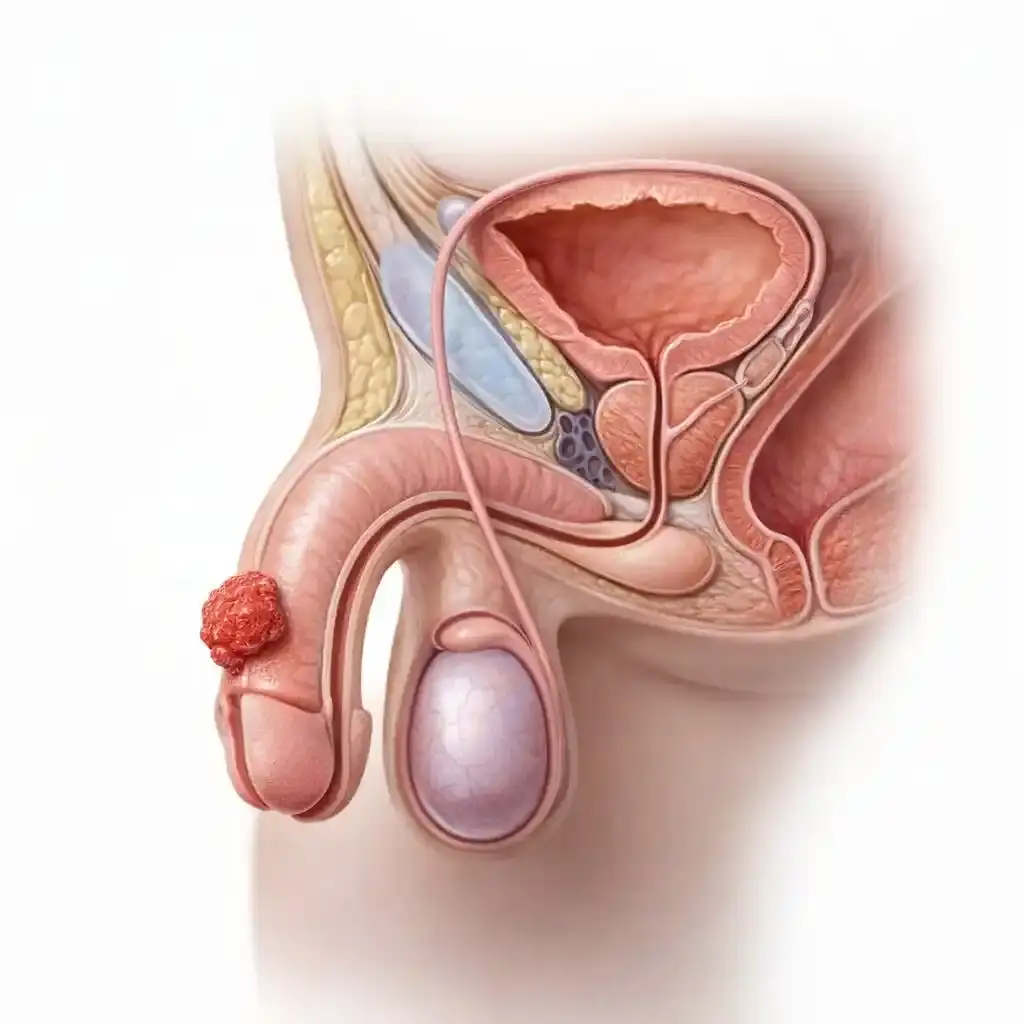
Penile cancer refers to the uncontrolled proliferation of cells in the penile tissue, similar to other cancers. The cancerous penile tissue then spreads to other parts of the body through various means. Penile cancer is generally rare, particularly in uncircumcised populations. It is diagnosed in less than 1 in 100,000 men and accounts for less than 1% of all cancers in men. Penile cancer is much more common in uncircumcised populations in parts of Asia, Africa, and South America.
What is penile cancer?
Penile cancer refers to the uncontrolled proliferation of cells in the penile tissue, similar to other cancers. The cancerous penile tissue then spreads to other parts of the body through various means. Penile cancer is generally rare, particularly in uncircumcised populations. It is diagnosed in less than 1 in 100,000 men and accounts for less than 1% of all cancers in men. Penile cancer is much more common in uncircumcised populations in parts of Asia, Africa, and South America.
The penis is the male external genitalia. It is also part of the urethra. Its tissue structure consists of skin, nerves, muscles, and blood vessels. The main part of the penis is known as the shaft, and the head of the penis is called the glans. At birth, the glans is covered by a piece of skin called the foreskin or prepuce. In male infants, the foreskin is usually removed in a surgical procedure called circumcision.
The penis contains three chambers within a soft, spongy network of blood vessels. Two of these cylindrical chambers, known as the corpora cavernosa, are located side-by-side on the upper part of the penis. The third cylindrical chamber is located beneath the other two and is known as the corpus spongiosum. The corpus spongiosum surrounds the urethra, a thin tube that originates in the bladder and passes through the penis. It mediates the passage of urine and semen and exits the body through an opening in the glans penis called the meatus—the external orifice.
Erection begins with a signal from the nervous system causing blood to fill the adjacent cylindrical structures and the opening to close. As this blood flow fills the cylindrical chambers, the spongy tissue expands, and the penis hardens and lengthens. During ejaculation, semen (sperm cells and fluids) flows through the urethra and is expelled from the body through the meatus (external orifice). After ejaculation, the valves open, blood flow decreases, and the penis becomes flaccid again.
Benign Lesions of the Penis
Benign penile lesions refer to non-cancerous, abnormal cell growths on the penis. These lesions typically appear as warts or irritated skin patches. As with penile cancer, they are most commonly found on the glans or foreskin, but less frequently can occur along the shaft of the penis.
1- Condylomas (genital warts)
These lesions can vary in size from tiny, cauliflower-like lesions visible only with a magnifying glass to giant lesions. These genital warts, called condylomas, are caused by sexually transmitted HPV (human papillomavirus) infections.
2- Bowenoid papüloz
This condition is also linked to HPV infection and tends to occur in younger, more sexually active men. It appears as small, red or brown spots or plaques on the penis shaft. These may resemble genital warts, but under a microscope they appear as dysplastic (abnormal) cells in the surface layer of the penis skin.
Bowenoid papulosis can be confused with an early-stage penile cancer called carcinoma in situ (CIS), also known as Bowen's disease. Often, bowenoid papulosis doesn't cause any problems and may even resolve on its own within a few months. However, if it doesn't resolve and is left untreated, it can, albeit rarely, progress to Bowen's disease.
Types of penile cancer
Various types of penile cancer can develop, originating from different tissues in the penis. Identifying these different types is crucial for determining the course of the disease and treatment methods. Almost all penile cancers begin in the skin cells of the penis.
1- Squamous cell carcinoma (Epidermoid Carcinoma)
About 95% of penile cancers originate from skin cells called squamous (epidermal) cells. Squamous cell carcinoma can start anywhere on the penis, but most often begins in the foreskin (in uncircumcised men) or the glans penis. These tumors tend to grow slowly and, if detected early, are usually treatable.
Verrucous carcinoma: Also known as Buschke-Lowenstein tumor, penile verrucous cancer is a rare type of squamous cell cancer that can begin in many areas of the penile skin. This cancer looks like a large genital wart, and they tend to grow slowly, although sometimes there are types that grow very quickly. While they can grow deep into the penile tissue, they rarely spread to other parts of the body, meaning they metastasize.
Carcinoma in situ (CIS): Carcinoma in situ (CIS) is the earliest stage of squamous cell carcinoma of the penis. At this stage, cancer cells are only found in the upper layers of the skin. They have not yet penetrated into deeper tissues.
Depending on where on the penis carcinoma in situ is located, other names are used for this disease.
- Glans carcinoma in situ lesions are called Queyrat erythroplasia.
- Carcinoma in situ lesions on the penile shaft or other parts are called Bowen's disease.
2- Melanoma
Melanoma is a type of skin cancer that begins in melanocytes, cells that help protect the skin from the sun and turn it brown. These cancers have a malignant prognosis, tending to grow and spread rapidly. They are more dangerous than the more common basal and squamous cell carcinomas of the penis. Melanomas are most often found in areas of the skin exposed to the sun, although rarely they can occur in other areas such as the penis. Only a very small percentage of penile cancers are malignant melanomas.
3- Basal cell carcinoma
Basal cell carcinoma is another type of skin cancer that can develop on the penis. It makes up only a small percentage of penile cancers. This type of cancer grows slowly and rarely spreads to other parts of the body, meaning it metastasizes.
4- Adenocarcinoma (Paget's disease of the penis)
This is a very rare type of penile cancer, originating from the sweat glands in the skin of the penis. It can be classified as carcinoma in situ (CIS) or adenocarcinoma (Paget's disease of the penis).
Separating them can sometimes be very difficult.
5- Sarcoma
The least common type of penile cancer is sarcomas. These cancers develop from blood vessels, smooth muscle, or other connective tissue cells of the penis.
Causes and Risk Factors of Penile Cancer
A risk factor can be defined as anything that affects your chances of developing a disease, such as cancer. Different cancers have different risk factors. Alongside some modifiable cancer risk factors, such as smoking or sun exposure, there are also non-modifiable risk factors, such as a person's age or family history.
Having certain risk factors, or even many, does not necessarily mean you will get penile cancer; penile cancer can occur even without any risk factors.
The identified risk factors for penile cancer are as follows.
1- Human Papilloma Virus (HPV) Infection
Human papillomavirus (HPV) is a DNA virus with nearly 200 types, about 60 of which infect humans, making it the most common sexually transmitted viral infection worldwide. In men, it most commonly manifests as genital warts (condylomas), and it is implicated in many cancers in both men and women, with approximately 15 types known to be carcinogenic. Different HPV types cause different types of warts in various parts of the body. HPV infection is known to cause cervical cancer, as well as cancers of the penis, tongue, oro-nasopharynx, larynx, and tonsils, and these can appear years after infection.
Certain HPV types have been associated with specific cancers and appear to be a significant risk factor for penile cancer. HPV traces are found in approximately half of penile cancers. It is detected in 70-100% of early-stage penile cancer tissues and 30-40% of advanced penile cancer tissues. Two proteins (E6 and E7) produced by high-risk human papillomavirus (HPV) types have been found to inhibit the function of tumor suppressor gene products in cells, causing these cells to become uncontrolled.
HPV infection is transmitted from one person to another through skin-to-skin contact, especially during sexual intercourse. HPV can be transmitted during many sexual activities, including vaginal, anal, and oral, but sexual contact is not necessary for infection to spread; skin-to-skin contact with an HPV-infected area of the body is sufficient for transmission. HPV infection can spread from one area of the body to another. For example, infection and lesions on the penis can spread to the anus over time.
HPV infection is very common worldwide, with half the world's population encountering the virus, and in 70% of cases, the body can clear the infection on its own. However, in some people with weakened immune systems, the infection cannot be cleared and becomes chronic. Over time, chronic infection, especially with certain HPV types, can lead to some types of cancer, including penile cancer. Uncircumcised men are more likely to become infected with HPV, develop chronic infection, and not be cleared.
See our HPV section to learn more.
2- Not being circumcised
At birth, the glans penis is covered by a piece of skin called the foreskin or prepuce. In male infants, the foreskin is usually removed in a procedure called circumcision.
Circumcision is usually performed on newborns, but it can also be done at later ages. Males who were not circumcised as children have a three times higher risk of penile cancer, and circumcision also reduces the risk of HPV infection. In fact, some publications claim that circumcision performed in childhood prevents penile cancer. The same protective effect is not seen to the same extent when circumcision is performed in adulthood.
While the exact reason for the lower risk in circumcised men is not entirely clear, it may be related to other known risk factors. For example, phimosis, a condition where the foreskin cannot be retracted due to narrowing, and smegma, the accumulation of secretory material under the foreskin, do not occur in circumcised men. Men with smegma or phimosis have a higher risk of penile cancer. The longer circumcision is delayed, the greater the likelihood of these risk factors developing.
When evaluating the risks and benefits of circumcision, it's important to consider the fact that penile cancer is very rare, even among uncircumcised men in America. While the American Academy of Pediatrics states that the health benefits of circumcision in newborn boys outweigh the risks, they also note that there isn't enough evidence to recommend routine circumcision for all newborns due to its benefits alone. Ultimately, decisions regarding circumcision are highly personal and often depend more on social and religious factors than on medical evidence.
3- Phimosis and Smegma
Phimosis: In uncircumcised men, this is a condition where the foreskin cannot be retracted due to adhesions or a narrow tip. The risk of penile cancer increases tenfold in the presence of phimosis. The reason for this is unclear, but it may be related to the accumulation of secretory material under the foreskin, known as smegma, and chronic inflammation associated with phimosis.
Smegma: The secretion that forms under the foreskin is called smegma. If this area is not cleaned regularly and properly, or if this secretion accumulates due to phimosis, it turns into a thick, sometimes foul-smelling substance called smegma. It is known that there is insufficient evidence to support claims that smegma contains substances that may cause cancer. It is believed that the irritation and infection caused by smegma, rather than smegma itself, do not cause penile cancer.
4- Use of Cigarettes and Other Tobacco Products
Men who smoke and/or use other types of tobacco have been found to be five times more likely to develop penile cancer. This risk is even higher for smokers and tobacco users who have an HPV infection. Smoking exposes the body to many cancer-causing chemicals (carcinogens). These substances can cause cancer in many different areas of the body through the bloodstream. It is stated that these substances lead to penile cancer by damaging genes in penile cells. DNA damage can lead to cancer when it affects genes that control cell growth.
5- UV light therapy for psoriasis
Some patients with a skin condition called psoriasis are treated with medications called 'psoralen' followed by ultraviolet-A (UVA) light therapy. This is called PUVA therapy. It has been found that men who undergo this treatment have a higher rate of penile cancer (9-10 times higher). To reduce this risk, the genitals of men treated with PUVA are now covered during the treatment.
6- Years old
The risk of developing penile cancer increases with age, and the average age of diagnosis is around 68. Approximately 4 out of 5 penile cancers are diagnosed in men over 55.
7- AIDS
Men with AIDS have a higher risk of penile cancer. This increased risk has been linked to the weakened immune system, a consequence of AIDS. However, it may also be linked to the presence of other risk factors in men with HIV (the virus that causes AIDS). For example, men with HIV are more likely to smoke and be infected with HPV.
8- Polygamy
Men who have had multiple sexual partners simultaneously or at different times, and who had their first sexual experience at a very young age, have been found to have a 3 to 5 times higher risk of developing penile cancer.
9- Poor Hygiene Conditions and Chronic Infections
Chronic infection and inflammation in the penis significantly increase the risk of penile cancer. The risk of developing penile cancer is much higher due to both sexually transmitted diseases and frequent penile infections (balanitis, postitis).
Symptoms of Penile Cancer
The symptoms and signs seen in penile cancer are not always specific to penile cancer; in fact, they often occur in conditions other than penile cancer. The possibility of penile cancer should be considered if the following symptoms and signs are present. The earlier the diagnosis, the earlier treatment can begin and the more successful it will be. The primary symptom of penile cancer is a persistent, stubborn, bleeding, or discharging sore on the penis that does not heal. Initially, it is mistaken for an infection and treated, but if it does not respond to treatment, cancer is suspected. Other symptoms include raised, wart-like redness, sores or hard lumps on the skin, itching and burning sensations, discharge, and palpable swelling in the groin area.
Skin changes: The first sign of penile cancer is certain changes seen in the skin of the penis. These are most often seen on the glans or foreskin of the penis, and less frequently on the shaft of the penis.
These changes are as follows:
- Thickening of the skin on the penis in some areas.
- Some changes in skin color
- A raised, wart-like sore or lump.
- A bleeding ulcerated wound (painful)
- A reddish, velvety rash under the foreskin.
- Small, hard-shelled lumps and swellings
- Flat, bluish-brown lesions/mass
- Foul-smelling discharge (fluid) or bleeding from under the foreskin
Sores or lumps caused by penile cancer often do not cause pain or other symptoms. Even without pain, it is advisable to consult a doctor if you notice any growth, swelling, sores, discharge, skin discoloration, or other abnormalities on your penis. Any changes in your penis that do not improve or worsen within four weeks should be checked by a doctor.
Swelling and Lumps in the Penis: A raised, wart-like sore or lump on the penis, especially in those with phimosis (narrowing of the foreskin), may be a sign of penile cancer.
Lumps under the skin in the groin area: Penile cancer often spreads first to the lymph nodes in the groin, and then to the pelvic lymph nodes further inside. Lymph nodes are considered part of the immune system. They are normally bean-sized and almost imperceptible; if there is a growth, it can be felt as smooth lumps under the skin. Enlarged lymph nodes do not always mean that cancer has spread there, as lymph nodes are more likely to enlarge in response to an infection. The tissue of penile cancer and the surrounding skin often become inflamed, which can cause nearby lymph nodes to swell and enlarge.
Diagnosis of Penile Cancer
Medical history and physical examination:Medical and sexual history and physical examination constitute the first step in diagnosing penile cancer. The medical history provides details such as when the symptoms started and whether they have changed. It also records any possible risk factors for penile cancer.
A physical examination should include a careful examination of the genital area for possible signs of penile cancer or other health problems. Lesions (sores) on the penis are carefully examined, and any lesions such as phimosis and HPV are checked. Enlarged lymph nodes in the groin are also palpated. If the symptoms and findings from the physical examination point to penile cancer, a biopsy of the lesion is necessary to confirm the diagnosis.
Biopsy:The only definitive way to determine whether a lesion on the penis is penile cancer is to perform a biopsy. For this, a small piece of tissue is taken from the lesion (wound) and sent to a pathology laboratory. The pathological examination will report whether or not the tissue contains cancer cells.
Types of biopsies and biopsy sites used in penile cancer.
- Incisional biopsy:Incisional biopsy refers to taking only a sample of the lesion and sending it to a pathology laboratory. In this type of biopsy, the lesion is not completely removed; only a sample is taken for diagnosis.
- Excisional biopsyIn excisional biopsy, the entire lesion (wound) is removed. This type of biopsy is most often used if the lesion is small, in the form of a nodule (lump) or plaque (raised, flat area).
- Lymph node biopsy:If the cancer has spread deep into the penis, the lymph nodes in the groin will need to be checked for cancer spread. These lymph nodes can be checked either by fine-needle aspiration or by surgical removal. This provides us with very valuable information in staging the disease.
- Fine needle aspiration (FNA):To perform this type of biopsy, a thin, hollow needle is inserted into the lymph node, and a few drops of fluid are aspirated and sent to pathology. If the enlarged lymph node is too deep to be felt by hand, access may be achieved with the aid of a CT scan or ultrasound.
- Surgical biopsy:In some cases, instead of fine-needle biopsy, lymph nodes in areas where cancer is likely to have spread are surgically removed. Removing and separately evaluating the sentinel lymph node, where the cancer first spread, is important in staging and treating penile cancer.
Computed tomography (CT):A CT scan can show how large a tumor is and can also help determine if the cancer has spread to the lymph nodes or other parts of the body.
CT-guided needle biopsy:CT scans can be used to guide a needle to take a biopsy from an enlarged lymph node or other areas where cancer may have spread.
Magnetic resonance imaging (MRI):MRI is a method used to identify areas where a tumor is likely to have spread, to determine if it has spread to other organs, and to stage the tumor.
Ultrasound:It can be useful for determining how deep the cancer has spread in the penis. It can also be used to visualize enlarged lymph nodes in the groin. Furthermore, it can serve as a guide for needle biopsies from various areas.
Chest X-ray: A simple chest X-ray can help check if the cancer has spread to the lungs.
Staging and Grading of Penile Cancer
After a diagnosis of penile cancer, the process of determining whether and how far the cancer has spread is called staging. Staging reveals the extent of the cancer's spread, and treatment strategies and methods may differ at each stage. Staging also helps predict the course of the disease and survival time. As a rule, the lower the number, the less the cancer has spread. A higher stage, such as stage IV, means the cancer has spread further. The earliest stage of penile cancer is stage 0, meaning the cancer has not spread beyond the top layer of skin. Other stages are I through IV. The staging and grading given below are used for squamous cell carcinoma, the most common type of penile cancer. Other types of cancer that originate in the penis, such as melanoma and sarcoma, are much less common and are staged using different systems.
How are the stages determined and what do they represent?
The most commonly used staging system for penile cancer is the American Joint Committee on Cancer (AJCC)-January 2018 TNM system, which is based on three key pieces of information:
Size of the main tumor (T):This indicates the extent to which cancer has progressed in the penis and whether it has spread to neighboring organs.
Spread to nearby lymph nodes (N):This indicates whether cancer has spread to regional lymph nodes, and if so, to what extent and in how many lymph nodes.
Spread to distant areas (metastasis) (M):This indicates whether the cancer has spread to distant lymph nodes or other parts of the body, such as the lungs, liver, or bones.
Grading of Penile Cancer
Another factor that can affect the stage of some cancers is the degree of aggressiveness of the cancer cells. This is a measure of how different cancer cells look from normal cells. The grade is usually indicated by a number from 1 to 3. The higher the number, the more abnormal the cells appear. High-grade cancers tend to grow and spread faster than low-grade cancers.
Penile cancer is classified as Grade (D) as follows:
- D-1: Well-differentiated, meaning relatively good.
- D-2: Moderately differentiated
- D-3: Poorly Differentiated
- D-4: Undifferentiated, severely differentiated.
After determining the TNM categories and grade of cancer, this information is combined with staging to determine a general stage. If the staging is based on information obtained from preoperative physical examination, biopsy, and imaging systems (CT scan, MRI, etc.), it is called clinical staging; if it is based on data obtained after pathological examinations, it is called pathological staging. The most accurate staging is pathological staging; the staging given below is pathological staging.
Stages of Penile Cancer: AJCC - January 2018
Stage 0: Tis or Ta, N0, M0
- Also called carcinoma in situ or CIS. The tumor is only in the upper layer of the skin and has not progressed deeper (Tis or Ta).
- The cancer has not spread to regional lymph nodes (N0) or to distant parts of the body (M0).
Hebrew I: T1a, N0, M0
- The tumor has spread to the tissue just beneath the top layer of skin. It has not spread to blood vessels, lymphatic vessels, or nerves in this area (T1a), and the cancer cells are not high-grade (not grade 3).
- The cancer has not spread to nearby lymph nodes (N0) or to distant parts of the body (M0).
Stage IIA: T1b, N0, M0; or T2, N0, M0
- The tumor has spread to the tissue just below the top layer of the skin, and has also spread to blood vessels, lymph vessels, or nerves in this area (T1b) and/or is high-grade (grade 3 and above).
- The cancer has not spread to regional lymph nodes (N0) or to distant parts of the body (M0).
Or
- The cancer has grown into the corpus spongiosum (the inner chamber that extends from the bottom to the tip of the penis).
- The cancer has not spread to nearby lymph nodes (N0) or to distant parts of the body (M0).
Stage IIB: T3, N0, M0
- The cancer has grown in the corpus cavernosum (two inner chambers that run along the upper part of the penis shaft).
- The cancer has not spread to nearby lymph nodes (N0) or to distant parts of the body (M0).
Stage IIIA: T1-3, N1, M0
- The tumor has grown in the tissue beneath the top layer of skin and may have grown into the corpus spongiosum and/or corpus cavernosum (T1 to T3).
- Cancer has spread to one or two regional inguinal lymph nodes on the same side of the body (N1). It has not spread to distant parts of the body (M0).
Stage IIIB: T1-3, N2, M0
- The tumor may have spread to the tissues beneath the top layer of skin and may have grown into the corpus spongiosum and/or corpus cavernosum (T1 to T3).
- Cancer has spread to 3 or more regional inguinal lymph nodes on the same side of the body, or to inguinal lymph nodes on both sides (N2).
- It has not spread to distant parts of the body (M0).
Stage IV: T4, Any N, M0; or Any T, N3, M0;
or Any T or Any N, M1
- The tumor has spread to nearby structures such as the scrotum, prostate, or pubic bone (T4).
- The cancer may or may not have spread to regional lymph nodes (any N). It has not spread to distant parts of the body (M0).
Or
- The tumor may or may not have grown into deeper layers of the penis or nearby structures (any T-shaped structures).
- The cancer has spread to regional lymph nodes in the pelvis or has grown outside of a lymph node and into the surrounding tissue (N3). The cancer has not spread to distant parts of the body (M0).
Or
- The tumor may or may not have grown into deeper layers of the penis or regional structures (any T). The cancer may or may not have spread to nearby lymph nodes (any N). The cancer has spread to distant parts of the body (M1).
Survival Rates for Penile Cancer
Survival rates give an idea of what percentage of people with the same type and stage of cancer are still alive for a certain period (usually 5 years) after diagnosis. These statistics can be confusing. They help us better understand the likelihood of successful treatment rather than how long the patient will live. When a 5-year survival rate of 80% is stated, it means that, on average, 80% of men with this cancer have a chance, or 80% of these patients are still alive after 5 years. These figures apply to the stage of the cancer at the time of initial diagnosis. These numbers do not account for everything. Survival rates are grouped according to how far the cancer has spread, but the patient's age, overall health, how well the cancer responds to treatment, and other factors also affect these rates.
When reporting survival rates for patients, the classification of cancer as localized, regional, or distant metastatic is considered, rather than the stage of the disease.
- Localized Disease: The cancer is confined to the penis.
- Regional Disease: The cancer has spread outside the penis to nearby structures or regional lymph nodes.
- Distant metastasis: Cancer has spread to distant parts of the body, such as the lungs, liver, or bones.
The 5-year relative survival rates for penile cancer are as follows:
- Localized-regional disease: 80%
- Regional Disease: 50%
- Disease with distant metastases: 9%
- From total combined grouping: 65%
Treatment of Penile Cancer
A multidisciplinary approach in the treatment of penile cancer.
Decision-making in cancer treatment requires combining a large amount of data, including symptoms and imaging information such as MRI/CT scans and PET scans, and necessitates a multidisciplinary approach—integration of all relevant departments—to select the most appropriate treatment for the patient. International standards in cancer treatment require this approach.
At Prof. Dr. Hakkı Perk's private clinic, treatment planning for uro-oncological, or cancer, patients is carried out using a multidisciplinary approach. Once a week, at a hospital with which I have an agreement, cancer patients are discussed in an oncological council attended by urologists, medical oncologists, radiation oncology specialists, nuclear medicine specialists, pathologists, and radiologists. Treatment decisions and processes for these patients are finalized. Treatment processes for patients with prostate cancer, kidney cancer, adrenal gland tumors, bladder cancer, and testicular cancer are determined with the participation of all relevant departments.
Furthermore, cancer patients are guided by international oncology guidelines such as NCCN, ASCO, EAU, and AUA in their treatment management. In such multidisciplinary collaborations and cancer councils, all aspects of a patient's condition are considered and decided upon. Feedback presentations regarding patients undergoing treatment are also given at these councils.
1-Surgical Treatment of Penile Cancer
Surgery is the most effective treatment for all stages of penile cancer. If the cancer is small and has not spread, it can usually be treated without having to remove part of the penis. If the cancer has progressed to a more advanced stage, it may be necessary to remove part or all of the penis along with the tumor. In patients with tumors that have grown deep into the penis (stage T2 or higher), it may be necessary to remove and sample some of the nearby lymph nodes in the groin. Instead of removing all the lymph nodes in the groin, the sentinel lymph node, where the cancer first arrived, may be removed, and if metastasis is present, then a wider range of lymph nodes may be removed.
Many different types of surgery are used in the treatment of penile cancer. Penis-preserving techniques are used as often as possible. Local treatments and limited surgeries are surgical choices made to preserve as much of the penis as possible in order to preserve sexual function, the appearance of the penis, and the ability to urinate standing up.
Circumcision:If the cancer is only in the foreskin, circumcision is often sufficient to treat the cancer. During this procedure, the foreskin and some skin appendages are removed, extending beyond the circumcision itself. If radiation therapy to the penis is planned, the radiation can cause swelling and tightening of the foreskin, which can lead to other problems, so circumcision is usually performed before this procedure.
Simple excisionIn simple excision surgery, the tumor is completely removed along with some nearby normal skin and tissue. If the tumor is small, the remaining skin can be sewn back on. This is done in the same way as an excisional biopsy.
Wide ExcisionIn a wide local excision, the tumor is removed along with a large amount of normal tissue surrounding it. Removing it with some healthy tissue reduces the likelihood of any cancer cells being left behind. If there is not enough skin left to cover the area, a skin graft can be taken from another part of the body and used on the area.
Mohs surgery (microsurgery - microscope-controlled surgery):
In some selected patients, it may be an option instead of wide local excision. In the Mohs technique, the layer of skin that the tumor may have invaded is removed, and then the sample is immediately examined under a microscope. If the removed tissue still contains cancer cells, another layer is removed and examined. This process is continued until no cancer cells remain in the skin sample.
This process is long and slow, but it means that more normal tissue can be saved, increasing the chance of preserving the penis as much as possible. This technique can be used for carcinoma in situ (CIS), where the cancer is only in the upper layers of the skin, and for some early-stage cancers that have not spread deep into the penis.
Glansectomy (Partial or complete removal of the glans penis):If the tumor is small and located only on the glans penis (tip of the penis), part or all of the glans may be removed. Skin grafts can be used to reconstruct the glans penis after surgery.
Partial or complete removal of the penis (penectomy):This operation removes part or all of the penis. This is the most common and best-known way to treat penile cancer that grows deep within the penis. The goal is to remove all the cancer. To do this, the surgeon must remove it along with a portion of the penis that appears normal.
If enough of the penis shaft (at least 2-3 cm) cannot be salvaged to allow the patient to urinate while standing, total penectomy is necessary. This means removing the entire penis, including the root portion extending into the pelvis. In this case, the urethra is anastomosed to the perineum, the area between the scrotum (testicle sac) and the anus, allowing the patient to urinate while sitting. This is called perineal urethrostomy. Because the sphincter muscle ("open-close" valve) in the urethra remains, the patient does not experience urinary incontinence and can control their urination.
For very advanced tumors, sometimes the penis is removed along with the scrotum (and testicles). This procedure is called 'emasculation'. Because this operation removes the testicles, which are the main source of the body's male hormone, lifelong male hormone supplementation is required after the procedure.
Lymph node surgery (lymphadenectomy):In patients with cancer that has grown deep within the penis (stage T2 or higher), it is often necessary to remove some nearby lymph nodes in the groin to check for cancer spread. Groin lymph nodes allow lymphatic fluid to return from the legs to the bloodstream. Removing many lymph nodes in this area can lead to problems with this drainage and cause abnormal swelling. This condition is called lymphedema. It is the most significant complication to expect in this surgery.
Sentinel lymph node biopsy (SLNB):Instead of removing all lymph nodes in this operation, only the sentinel lymph node, where cancer is most likely to spread first, is removed. If there is no cancer in this node, further intervention is avoided; if cancer is present, a more comprehensive operation known as inguinal lymphadenectomy is performed. This method results in fewer problems with lymphedema and wound healing after lymph node removal. The disadvantage of this method is that metastases beyond the sentinel lymph node may be missed.
Inguinal lymphadenectomy (groin lymph node dissection):Many patients with penile cancer have enlarged inguinal lymph nodes detected at the time of initial diagnosis. These lymph node enlargements may simply be due to existing inflammation and can be resolved with antibiotics. If these enlargements do not disappear or shrink sufficiently with antibiotics, the lymph nodes are surgically removed (inguinal lymphadenectomy). If the lymph nodes are palpable, a lymph node biopsy may be recommended in cases of high-stage or advanced cancer.
Pelvic lymph node surgery:If cancer is found in two or more inguinal (groin) lymph nodes, the pelvic lymph nodes should also be removed and examined. This can be done in the same procedure or as a separate operation later.
2- Radiation Therapy (Radiotherapy) for Penile Cancer
Radiation therapy uses high-energy beams or particles to destroy cancer cells. In uncircumcised patients who will receive radiation to the penis, circumcision is performed beforehand because radiation therapy can cause swelling and compression of the foreskin. There are two main ways to administer radiation therapy for penile cancer: external radiation and brachytherapy (internal).
In penile cancer, radiotherapy is used in the following situations:
- For some small penile cancer lesions, radiotherapy may be an option that does not require surgery.
- If cancer has spread to the lymph nodes, radiotherapy may be used after lymph nodes are removed to reduce the likelihood of recurrence.
- For patients who do not want surgery or for whom surgery is not considered an option, radiotherapy can be used as a treatment plan.
- Radiotherapy can also be used in patients with advanced cancer to slow the growth of cancer or to relieve the symptoms it causes.
- To shrink the tumor and make it easier to remove it with less damage to the penis, radiotherapy may be given in combination with chemotherapy before surgery.
- In patients at high risk of metastasis to regional lymph nodes, radiotherapy may be added to existing treatment to reduce this risk.
External radiation therapy:The most common way to deliver radiation therapy is with focused beams of radiation from an external device that targets the tumor. This type of radiotherapy is usually given in a fixed number of sessions, 5 days a week, for 6 weeks.
A wax or plastic block or mold can be used to hold the penis in the exact same position during treatment. Shields can also be used to protect the groin and testicles.
Brakiterapi:Brachytherapy involves placing a radioactive seed into tumor tissue. The implanted radioactive substance emits radiation over time, treating the tumor while minimizing damage to healthy tissue and eliminating the need for hospitalization.
There are two ways to receive brachytherapy for penile cancer.
- Interstitial radiation:In this method, hollow needles are inserted into the penile tumor under operating room conditions. The needles are left in place for several days. Small seeds of radioactive material are released from these needles several times a day. After the treatment is complete, all the seeds and needles are removed.
- Plesiobrachytherapy:In this type of brachytherapy, the radiation source is placed near the tumor, not directly on it. A plastic cylinder is then placed around the penis. Another cylinder, holding the radiation source, is then placed on top of the first. Treatment is usually continued for several days in a row.
Possible side effects of radiation therapy include:The main disadvantage of radiation therapy is that it can damage healthy tissue surrounding cancer cells. The skin in the treated area usually becomes red and sensitive, may peel, and swelling may occur in the radiation area. It may cause a burning sensation when urinating for a while. The penis may lose some elasticity due to radiation therapy, and localized vasodilation (telangiectasia) may occur. Hygiene and skin care are key to preventing inflammation in the area. Fatigue, nausea, and diarrhea may sometimes occur after radiotherapy to the pelvic region and inguinal lymph nodes.
In patients treated with brachytherapy, side effects begin to appear approximately 3 weeks after treatment, and recovery takes about 12 weeks. In external radiotherapy, side effects begin gradually during treatment and improve over time after treatment ends. Most disappear within a few months.
Less common, but more serious side effects include:
- Some of the skin or tissue at the tip of the penis may die (a condition called necrosis).
- Urethral stricture may develop, which can make urination difficult.
- A fistula, or hole, can occur between the urethra and the skin.
- Radiation therapy to the shaft of the penis can cause erectile dysfunction. Radiotherapy to the tip of the penis (glans) has little effect on erection.
3- Local Treatments (Non-Surgical) for Penile Cancer
Some very early-stage, low-grade penile cancers, particularly carcinoma in situ (where the cancer has only affected the upper layers of the skin), can be treated with non-surgical techniques. These include radiation therapy, laser ablation, cryotherapy, and certain locally applied cancer drugs. These types of treatments can be called penis-sparing techniques. These treatments cause less damage to the integrity and structure of the penis. Of course, it should be kept in mind that these local treatments should be used in relatively low-grade tumors that have not spread, and are not suitable for cancers that have spread or have a high potential for spreading.
Laser ablation:The use of laser beams can be beneficial for squamous cell carcinoma in situ (CIS) and very thin or shallow basal cell carcinomas. It can also be offered as an option for patients with low-stage tumors who refuse surgery. The procedure is performed after local anesthesia; healing of the laser wound may take some time, and the procedure can be repeated if local tumor recurrence occurs.
Kriyocerrahi-Kriyoterapi:Cryosurgery is performed in almost the same way as laser surgery, using liquid nitrogen to freeze and kill cancer cells. It is used for some verrucous penile cancers and superficial cancer of the glans penis (CIS). After the area is locally anesthetized, cryotherapy is applied, freezing the area, and the resulting wound on the skin takes several months to heal.
Use of local cancer drugs (topical treatments): In topical chemotherapy, certain cancer drugs are administered orally, by intravenous injection, or applied as a cream to the affected skin.
- 5-Uracil (5-FU):The most commonly used cancer drug for this purpose is 5-fluorouracil (5-FU). It is applied topically to the cancerous skin for several weeks and kills cancer cells in the upper layers of the skin, but it is not used in patients with cancer that has grown deep within the skin or spread to other organs. Therefore, treatment with 5-FU is mostly used in precancerous lesions or superficial cancers called carcinoma in situ (CIS). Redness and tenderness may occur on the skin for a few weeks after application.
- Otherwise:It is used as a topical cream for superficial penile cancer. It causes the immune system to react against the cancer and destroy it. It is applied to the skin approximately 3 to 7 times a week for several weeks. It can cause skin irritation, which may be severe in some patients.
Photodynamic therapy (PDT):PDT (Percutaneous Tetralogy) is not a very commonly used method for penile cancer; it may only be offered as an option in certain cases. This treatment uses special medications and laser light for cancers near the surface of the penis. The medications used in conjunction with the laser aim to pinpoint the areas where the laser is applied and increase precision.
4- Systemic Chemotherapy in Penile Cancer
Chemotherapy refers to the classic systemic chemotherapy regimens used in penile cancer. Systemic chemotherapy uses cancer drugs administered intravenously or orally. This treatment is most commonly used for penile cancers that have spread to lymph nodes or distant organs. Chemotherapy can also be administered to shrink tumors before surgery to make them easier to remove (called neoadjuvant chemotherapy) or after surgery (called adjuvant chemotherapy), and it can also be used to prevent cancer recurrence. Chemotherapy is given in cycles—courses—and a rest period is placed after each treatment cycle to allow time for the body to recover. Chemo cycles usually last about 3 to 4 weeks.
The chemotherapy drugs used in penile cancer are as follows:
- Pilate
- Uracil (5-FU)
- Paclitaxel (Taxol®)
- İfosfamid (Ifex®)
- MTX
- Kapesitabin (Xeloda®)
These drugs are usually given in combinations of two or three. They are used in cases of penile cancer that has spread to lymph nodes or other organs.
The most common drug combinations include: combinations that contain:
- Pilatin-5-FU
- TIP: Paklitaksel (Taxol), ifosfamid and sisplatin ("platin")
Possible side effects of chemotherapy: Chemotherapy drugs are more effective against rapidly dividing cells, so they are effective against cancer cells, but they can also damage healthy cells and tissues. For example, cells in the bone marrow (where new blood cells are made), the mucous membranes (surface) of the mouth and intestines, and hair follicles also divide relatively quickly and can be affected by chemotherapy, leading to some side effects. The side effects of chemotherapy depend on the type of drugs, the dose, and how long they are used. These side effects usually disappear over time after treatment ends.
Common side effects may include:
- Hair loss
- Mouth sores
- Loss of appetite
- Nausea and vomiting
- Diarrhea or constipation
- Increased chance of infection (from low white blood cell count)
- Easy bruising or bleeding (from low blood platelet count)
- Fatigue (due to anemia)
The following are side effects specific to the chemotherapy drug used:
- Cisplatin and paclitaxel can cause nerve damage (neuropathy), which can lead to numbness and tingling in the hands and feet. Cisplatin can also cause kidney damage (nephropathy). To help prevent this, large amounts of intravenous (IV) fluids are given with cisplatin.
- 5-fluorouracil (5-FU) and capecitabine can cause sores (mucositis) in the mouth that can make eating difficult. These medications can also cause diarrhea.
- Ifosfamide can damage the inner lining of the bladder (hemorrhagic cystitis). To prevent this, a medication called Mesna is often given along with ifosfamide.
The information here is for general education only and is not medical advice. Please consult your physician for personalized treatment.