









Prostate Cancer
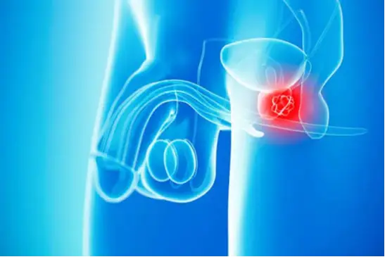
Prostate cancer is a malignant tumor that develops in the prostate and refers to uncontrolled cell growth. The prostate is a gland found only in men. It is an organ that produces part of the semen and secretes substances necessary for sexual and reproductive functions.
Prostate cancer is a malignant tumor that develops in the prostate and refers to uncontrolled cell growth. The prostate is a gland found only in men. It is an organ that produces part of the semen and secretes substances necessary for sexual and reproductive functions. The prostate is approximately the size of a chestnut, located below the bladder and surrounding the bladder outlet, and the urethra, which carries urine and semen out of the body through the penis, passes through the center of the prostate. As men age, the size of the prostate may change and enlarge; this may occur as benign prostate enlargement or in the direction of prostate cancer. The two conditions may coexist, but one does not transform into the other.
Prostate cancer is the most common type of cancer seen in men, and 1 in 8 men has a clinical possibility of developing prostate cancer during their lifetime. Prostate cancers that do not become clinically apparent and are not treated are 3-4 times more common. Developments in diagnostic methods and increased life expectancy have increased the number of diagnosed prostate cancer cases. Although it is so common, it is not a highly fatal cancer. As urologists, we can diagnose only 1/5 of all prostate cancers, and only 1/5 of the patients we diagnose and follow up die because of prostate cancer. Most prostate cancers grow slowly and remain limited to the prostate gland, and sometimes treatment is not required, whereas some other types are wild and aggressive and can spread rapidly. In general, the life expectancy of patients with prostate cancer is relatively long, and this period continues to increase with developments in diagnosis and treatment of malignant prostate cancer. I believe that asking the question “Is there a treatment for prostate cancer?” constitutes an answer after this information.
Types of Prostate Cancer
More than 95% of all prostate cancers consist of conventional acinar type adenocarcinoma. These cancers develop from the cells that produce prostate fluid. When prostate cancer is mentioned, prostate adenocarcinoma is understood.
Other types of cancer that can start in the prostate include:
- Small cell carcinomas
- Neuroendocrine tumors (except small cell carcinomas)
- Transitional epithelial cell carcinomas (such as bladder cancer)
- Sarcomas
Some prostate cancers grow and spread rapidly, but most grow slowly. In fact, autopsy studies have shown that many elderly men (and even some younger men) who died from other causes had prostate cancer that never affected them during their lives. In most cases, neither they nor their doctors even knew they had it.
Causes and Risk Factors of Prostate Cancer
Age
Aging is among the definite risk factors for prostate cancer, because as age increases, the risk of prostate cancer also increases. The average age of occurrence is 66, and 60% of cases are seen in people aged 65 and older, while it is very rare under the age of 40. The likelihood of developing prostate cancer rises rapidly after the age of 50.
Race and Ethnicity
Race and ethnicity are also among the definite risk factors, just like age. Prostate cancer develops more frequently in black men compared to men of other races and tends to occur at an earlier age. It is seen least frequently in East Asians. The reasons for these racial and ethnic differences are not clear.
Familial and Genetic Predisposition
Genetic and familial predisposition are also considered definite risk factors. Prostate cancer is seen more frequently in some families, suggesting that hereditary and/or genetic factors may be effective. However, family history is not detected in most prostate cancer cases. It is estimated that 9% of all prostate cancers and 45% of prostate cancers under the age of 55 are caused by familial and genetic predisposition, showing that familial and genetic prostate cancers tend to occur at earlier ages.
It is a known fact that a man whose father or especially brother has prostate cancer has more than double the risk of developing this disease. This demonstrates how important familial predisposition is as a risk factor. The risk is much higher in men who have several relatives affected by the disease, especially if the relatives were diagnosed at a young age.
Although some inherited gene mutations appear to increase the risk of prostate cancer, they probably account for only a small percentage of cases. Inherited mutations in the BRCA1 or BRCA2 genes, which are associated with an increased risk of breast and ovarian cancer in some families, have also been found to increase the risk of prostate cancer in men (especially BRCA2 mutations). Men with Lynch syndrome caused by inherited gene changes have also been found to carry a high risk for many cancers, including prostate cancer.
Geographic Region
Prostate cancer is most common in North America, northwestern Europe, Australia, and the Caribbean islands. It is less common in Asia, Africa, Central America, and South America. Although the reasons are not clear, it is thought that more intensive screening for prostate cancer in some developed countries and other factors such as lifestyle differences (diet, etc.) are effective. The frequency of the disease varies between 5-111 per 100,000 people depending on geographical regions.
Diet
Diet is among the possible risk factors. Although the exact role of diet in prostate cancer is not clear, men who consume too much milk and dairy products seem to have a slightly higher chance of developing prostate cancer. It has been suggested that men who consume too much calcium may have a higher risk of developing prostate cancer. However, there is no such connection with calcium levels found in a normal diet. It has been found that both lower than normal and higher than normal vitamin D levels increase the risk of prostate cancer (especially high-grade cancer). High alcohol consumption has been found to increase the risk of developing prostate cancer and the risk of cancer-related death. No relationship has been found between long-chain omega-3 unsaturated fatty acid intake and prostate cancer. However, fried food consumption is known to increase the risk. Findings regarding the reduction of prostate cancer risk with tomato intake (cooked) and lycopene have not been proven. There is also no evidence that red and processed meat consumption increases the risk of prostate cancer.
Obesity, Height, and Prostate Cancer Relationship
According to studies, height and weight may be associated with a man’s risk of developing prostate cancer and dying from this cancer. In a study designed to examine the relationship between nutrition, environmental factors, lifestyle, and cancer risk, it has been suggested that the likelihood of being diagnosed with prostate cancer increases as a man’s height and body size increase.
Every 10 cm increase in waist circumference was found to increase the risk of advanced prostate cancer by 13% and the likelihood of dying from the disease by 18%. For every 10 cm increase in height, the likelihood of men being diagnosed with high-grade prostate cancer increased by 21%, and the likelihood of dying from this disease increased by 17%. These results should not be misunderstood; rather than indicating that tall men have a higher risk of developing prostate cancer, they suggest that the risk of developing high-risk prostate cancer and dying from these cancers may be higher. Height is not associated with total prostate cancer risk or the development of slow-growing tumors, but it has been linked with an increased risk of being diagnosed with aggressive-poor prognosis prostate cancer and dying from this disease.
Weight and body mass index may play a much greater role in cancer risk. Men who are overweight and have a larger waist circumference are more likely to be diagnosed with aggressive-poor prognosis prostate cancer. This may be because obese men are less likely to be diagnosed with cancer or have their cancer detected early, and prostate-specific antigen (PSA) concentrations may appear lower in these men. Conversely, having a healthy body weight may be associated with a reduced risk of high-grade prostate cancer and a lower risk of death from prostate cancer years later.
Although there is nothing you can do to change your height, lifestyle factors can affect obesity and therefore cancer risk. The best thing to do to reduce the risk of prostate and other cancers is to maintain as healthy a lifestyle as possible. In particular, losing weight may reduce the risk, since waist circumference may be associated with aggressive cancer development. Making informed decisions may help you stay healthy or lead to early diagnosis, which may save your life if diagnosed.
Male Hormones
Androgens such as testosterone (male hormones) support the growth and proliferation of prostate cells, but their role in the formation of prostate cancer is not entirely clear. It has been found that the incidence of prostate cancer is below average in men with very low free testosterone concentrations (the lowest 10%). In addition, average testosterone levels were found to be 15–20% higher in the black population compared to other races, and the high incidence of prostate cancer in black men has been attempted to be explained by this, but this relationship has not been proven and has only been claimed as a potential risk factor. Having higher androgen levels may contribute to the risk of prostate cancer in some men. Tumors develop when cells begin to grow faster than normal. The growth of prostate cancer cells depends on male hormones known as androgens. The most important androgen hormone is testosterone, which is synthesized predominantly in the testicles.
Smoking
Although no direct connection has been found between smoking and prostate cancer, it has been determined that smokers have a higher risk of dying from prostate cancer.
Exposure to Chemicals
Exposure to chemicals is among the potential risk factors. There is some evidence that firefighters and people working night shifts may be exposed to chemicals that could increase the risk of prostate cancer. Occupational exposure to cadmium (Cd) has also been found to increase the risk of prostate cancer.
Sexually Transmitted Infections
Sexually transmitted infections are also among the potential risk factors. Researchers have found that sexually transmitted infections (such as gonorrhea or chlamydia) increase the risk of developing prostate cancer. It has also been determined that sexually transmitted HPV infection increases this risk.
Vasectomy
Vasectomy is also mentioned as a potential factor. Although it has been suggested that men who have undergone vasectomy (sterilization) may have a slightly higher risk of prostate cancer, this has not been fully proven.
Other Risk Factors
- Inflammatory bowel disease (Crohn’s disease, ulcerative colitis)
- Higher risk of death from prostate cancer has been found in men with hair loss (baldness)
- People positive for Human Papilloma Virus (HPV type 16) have been found to have a higher risk of prostate cancer
- The estrogenic insecticide chlordecone has been associated with an increased risk
- History of acne on the skin
- Ultraviolet rays have been claimed to increase the risk
- Higher ejaculation frequency: Ejaculating 20 times or more per month has been found to reduce the risk of prostate cancer by 20%
- Radioactive radiation
- Certain mold fungi on food products
How Does Prostate Cancer Develop?
How prostate cancer develops, as with other cancers, is not known with certainty. Prostate cancer is the uncontrolled growth and proliferation of cells in the prostate. Some genes that help cells grow, divide, and survive are called oncogenes. Genes that normally keep cell growth under control, repair mistakes in DNA, or cause cells to die at the correct time are called tumor suppressor genes. It is thought that DNA mutations (or other changes) that keep oncogenes active and/or deactivate tumor suppressor genes cause the development of prostate cancer. These types of gene changes (which may be inherited or acquired during life) are stated to cause cells to grow out of control. It is estimated that inherited gene changes play a role in approximately 10% of prostate cancers.
1- Inherited Gene Mutations (Changes)
BRCA1 and BRCA2
These belong to the tumor suppressor gene group. Inherited mutations in these genes are mostly known to cause breast and ovarian cancer in women, and they are also held responsible for the development of prostate cancer (especially BRCA2).
CHEK2, ATM, PALB2 and RAD51D
Mutations in these and other tumor suppressor DNA repair genes may also be responsible for some hereditary prostate cancers.
DNA mismatch repair genes (such as MSH2, MSH6, MLH1 and PMS2)
These genes normally help correct errors (mismatches) in DNA when a cell prepares to divide into two new cells. Men with inherited mutations in one of these genes (such as patients with Lynch syndrome) have an increased risk of prostate and certain other cancers.
RNASEL (formerly HPC1)
The normal function of this tumor suppressor gene is to help cells die when something goes wrong inside them. Inherited mutations in this gene may allow abnormal cells to survive longer than they should, which can lead to an increased risk of prostate cancer.
HOXB13
This gene is important in the development of the prostate gland. Mutations in this gene have been associated with early-onset prostate cancer seen in some families. Fortunately, this mutation is rare.
2- Acquired Gene Mutations
Some genes mutate during a person’s lifetime, and these mutations are not passed on to children. These changes are found only in cells derived from the original mutated cell. These are called acquired mutations. Most gene mutations associated with prostate cancer are acquired during a person’s life rather than inherited. How often these acquired DNA changes occur randomly and how often they are affected by other factors (diet, hormone levels, etc.) is not clear. In general, the faster prostate cells grow and divide, the greater the chance for mutations to occur. Therefore, anything that accelerates this process may make prostate cancer more likely.
Examples
- Androgens such as testosterone support prostate cell growth. Having higher androgen levels may contribute to the risk of prostate cancer in some men.
- Men with higher levels of another hormone called insulin-like growth factor-1 (IGF-1) have been found to have a higher likelihood of developing prostate cancer.
- Inflammation in the prostate may be associated with prostate cancer, and it has been suggested that inflammation can cause DNA damage in normal cells, contributing to their transformation into cancer cells.
- Exposure to radiation or cancer-causing chemicals can cause DNA mutations in many organs, but these have not been shown to be major causes of mutations in prostate cells.
What Are the Symptoms of Prostate Cancer?
Most prostate cancers are detected early through screening. Early prostate cancer usually does not cause any symptoms. Most symptoms that are seen are more likely to be caused by something other than prostate cancer. Difficulty urinating is much more often caused by non-cancerous enlargement of the prostate.
Prostate cancer may cause the following symptoms:
- Urination complaints including slow or weak urine flow or the need to urinate more frequently, especially at night (burning, frequent need to urinate, inability to completely empty the bladder)
- Blood in the urine or semen
- Erectile dysfunction (ED), which may occur in advanced stages
- Pain caused by cancer spreading to the hips, back (spine), chest (ribs), or other bones
- Weakness or numbness in the legs or feet, or even loss of bladder or bowel control due to cancer pressing on the spinal cord
Tests Performed to Diagnose and Stage Prostate Cancer
Most prostate cancers are found as a result of health screenings. Early prostate cancers usually do not cause symptoms, although they are sometimes detected because of the symptoms they cause; however, symptomatic prostate cancers are generally more advanced than asymptomatic ones. The definitive diagnosis of prostate cancer can only be made with a prostate biopsy. As with other cancers, a systematic step-by-step process is followed for the diagnosis of prostate cancer. In a patient suspected of having prostate cancer, medical history and physical examination constitute the first step of diagnosis. Questions are asked about urinary or sexual function problems, what symptoms are present, and how long they have been experienced. Questions may also be asked about possible risk factors, including family history. In the physical examination, digital rectal examination is essential, and the physician attempts to feel whether there is a nodule, lump, or hard area. It may also help determine the size and extent of suspicious areas and whether they are on one side of the prostate, both sides, or spread beyond the prostate into nearby tissues.
Prostate-Specific Antigen (PSA) Blood Test – What is PSA?
Prostate-specific antigen (PSA), known as the prostate blood test, is a protein produced by cells in the prostate (both normal and cancer cells). PSA is found in large amounts (a million times higher) in semen and is an enzyme that allows semen to liquefy in the external environment, while a small amount also passes into the bloodstream. PSA levels in the blood are measured in units of nanograms per milliliter (ng/mL). As PSA levels rise, the likelihood of prostate cancer increases, but there is no definite threshold that can conclusively determine whether prostate cancer is present. Most commonly, a PSA threshold value of 4 ng/mL or higher is used, but starting from a lower level such as 2.5 or 3 ng/mL may also be recommended.
Most men without prostate cancer have PSA levels below 4 ng/mL. When prostate cancer develops, PSA levels usually rise above 4. However, a PSA level below 4 does not guarantee the absence of cancer. Approximately 15% of men with PSA levels below 4 ng/mL may have prostate cancer if a biopsy is performed. The range between 4–10 ng/mL is called the gray zone or borderline range, and the approximate risk of prostate cancer in this range is 25%. If PSA is above 10, the likelihood of prostate cancer is over 50%.
Factors That Can Affect PSA Levels
One reason why it is difficult to use a strict PSA threshold while searching for prostate cancer is that many factors other than cancer can also affect PSA levels.
Factors That May Increase PSA Levels
1- Prostate Enlargement
Benign prostate enlargement (BPH), a non-cancerous enlargement affecting many men as they age, can increase PSA levels.
2- Advanced Age
PSA levels normally rise gradually with aging even without prostate problems.
3- Prostatitis
Prostate infection is an important clinical condition that can raise PSA levels, sometimes significantly in acute infections.
4- Ejaculation
Ejaculation may temporarily raise PSA levels. Therefore, avoiding ejaculation for one or two days before testing is recommended.
5- Cycling
Some studies have suggested that cycling may temporarily increase PSA levels, although this has not been proven.
6- Certain Urological Procedures
Procedures affecting the prostate, such as prostate biopsy or cystoscopy, may temporarily increase PSA levels. Some studies have suggested that digital rectal examination (DRE) may slightly raise PSA levels, although this has not been proven. As a rule, PSA testing is recommended before digital rectal examination.
7- Certain Medications
Male hormones such as testosterone and medications containing these hormones may increase PSA levels.
Factors That May Lower PSA Levels (Even If Prostate Cancer Exists)
1- 5-Alpha Reductase Inhibitors
Some medications used to treat benign prostate enlargement or urinary complaints, such as Dutasteride, reduce PSA levels by half within 3 months. In patients using these medications, PSA values are multiplied by two for interpretation. These drugs may also affect prostate cancer risk and may mask prostate cancer because they lower PSA.
2- Herbal Mixtures
Some mixtures sold as dietary supplements may mask elevated PSA levels. Saw palmetto, commonly used for benign prostate enlargement, does not appear to affect PSA significantly.
3- Some Other Medications
Some studies have suggested that long-term use of aspirin, statins (cholesterol-lowering drugs), and thiazide diuretics (such as hydrochlorothiazide) may lower PSA levels.
Special PSA Types
PSA is sometimes called total PSA because it includes different PSA forms. If PSA screening results are abnormal, additional PSA tests may help determine whether a prostate biopsy is necessary.
Percentage of Free PSA
PSA exists in two main forms in the blood. One form is bound to blood proteins, while the other circulates freely. The percentage of free PSA (%fPSA) indicates what percentage of total PSA is free PSA. The percentage of free PSA is lower in men with prostate cancer than in men without cancer. Normally it is expected to be above 25%, especially in patients whose total PSA level is in the gray zone range of 4–10 ng/mL, helping guide biopsy decisions.
Low free PSA and high total PSA levels increase the likelihood of prostate cancer and influence biopsy decisions. Prostate biopsy is recommended in patients whose free PSA percentage is 10% or lower, and biopsy should be considered if this ratio is between 10–25%. Using these ratios helps diagnose many cancers and avoid unnecessary biopsies.
Complex PSA
This test directly measures the amount of PSA bound to other proteins (the “non-free” portion of PSA). It can be used instead of total and free PSA and provides similar information, although it is not widely used.
Tests Combining Different PSA Types
Some newer tests combine results from different PSA types to generate an overall score reflecting the likelihood of prostate cancer.
These tests include:
- Prostate Health Index (PHI), combining total PSA, free PSA, and proPSA
- 4Kscore test, combining total PSA, free PSA, intact PSA, and human kallikrein 2 (hK2) with certain other factors
These tests may help determine whether a patient with mildly elevated PSA should undergo prostate biopsy. They can also help determine whether a repeat biopsy is necessary in patients whose previous biopsy did not detect cancer.
PSA Velocity
PSA velocity is not a separate test. It measures how quickly PSA rises over time. Normally, PSA levels increase slowly with age. In prostate cancer patients, PSA levels have been found to rise more rapidly. Although not widely used, an increase greater than 0.74 ng/mL per year may be considered significant.
PSA Density (PSAD)
The prostate gland volume is measured by transrectal ultrasound, and PSA is divided by prostate volume, giving the PSA amount per gram of tissue. The normal value is expected to be below 0.15, and biopsy may be considered if it exceeds 0.15. PSA density is not considered more useful than the percentage of free PSA.
Age-Specific PSA Ranges
Blood PSA levels are normally higher in older men than in younger men. A PSA level at the upper limit of normal may be concerning in a 50-year-old man but less concerning in an 80-year-old. Some age-specific PSA levels have been defined, although this is not commonly used.
In addition to being a very important marker used in the diagnosis of prostate cancer, PSA is also used in patients diagnosed with prostate cancer for staging, determining which imaging tests should be performed, treatment decisions, and treatment follow-up. Along with PSA, the tumor grade found in biopsy (Gleason score) also guides decisions about whether imaging studies such as CT, MRI, and bone scintigraphy should be performed. PSA testing is also an important part of monitoring treatment effectiveness and detecting possible recurrence after treatment.
Prostate Biopsy – How Is a Prostate Biopsy Performed?
If the results of the PSA blood test, digital rectal examination, or other tests indicate prostate cancer, a prostate biopsy will be necessary. A biopsy is a procedure in which small samples are taken from the prostate and then examined under a microscope. The main method used to diagnose prostate cancer is the standard systematic 12-core needle biopsy.
During the biopsy, the prostate is usually targeted using an imaging test such as transrectal ultrasound (TRUS), MRI, or a fusion of both. The doctor inserts a thin needle into the prostate either through the rectal wall (transrectal biopsy) or through the area between the scrotum and anus (transperineal biopsy). When the needle is withdrawn, small pieces of prostate tissue are placed into pathology containers. Most of the time, approximately 12 core samples are taken from different regions of the prostate.
Although the biopsy procedure may seem painful, each biopsy is generally comfortable because it is performed with a special biopsy device that works only briefly. The device inserts and removes the needle in less than a second. Before the biopsy, local anesthesia is injected around the prostate to numb the area. The biopsy takes approximately 10 minutes and is usually performed in the doctor’s office. Antibiotics are recommended before the biopsy and for 1–2 days afterward to reduce the risk of infection. After the procedure, slight pain in the area and blood in the urine may occur for several days. Mild rectal bleeding may also occur, especially in patients with hemorrhoids. Difficulty urinating or inability to urinate after prostate biopsy may occur. Blood in the semen or rust-colored semen that may last for several weeks can also be seen after the procedure.
Evaluation of Biopsy Results
Biopsy results may be reported as follows:
- Cancer positive: Cancer cells were seen in the biopsy samples.
- Cancer negative: No cancer cells were seen in the biopsy samples.
- Suspicious: Something abnormal was seen, but it may not be cancer.
If the Biopsy Is Negative
If prostate biopsy results are negative (meaning not cancer), it may still not mean there is no cancer, because the needle may have missed cancerous tissue. In the following process, regular PSA and digital rectal examination follow-ups are performed. The need for a second biopsy is decided according to the results of these follow-ups and other imaging tests such as multiparametric MRI (mpMRI), and the timing is planned accordingly.
If prostate cancer is still strongly suspected, the following may be recommended:
Other Laboratory Tests
Examples include:
- Prostate Health Index (PHI)
- 4Kscore test
- PCA3 tests (such as Progensa)
- ConfirmMDx
Repeat Prostate Biopsy
In addition to the standard systematic 12-core biopsy, extra samples may be taken from regions not biopsied initially, or targeted biopsy (fusion biopsy) may be performed on suspicious areas identified in multiparametric MRI images.
Prostate Cancer Grade (Gleason Score or Grade Group)
If prostate cancer is present in the pathological examination of the biopsy, it is graded from 1 to 5 according to how aggressive the cancer appears. The grade depends on how abnormal the cancer cells look under the microscope. Higher-grade cancers appear more abnormal and are more likely to grow and spread rapidly. This grading system is known as Gleason grading and is still actively used today.
1- Gleason Score
If the cancer closely resembles normal prostate tissue, it is called grade 1. If it appears very abnormal, it is reported as grade 5. Grades 2 to 4 have features between these two extremes. Nearly all prostate cancers are grade 3 or higher; grades 1 and 2 are rarely used.
Because prostate cancers often contain areas with different grades, the two most dominant grades are identified and added together to determine the Gleason score. The first grade represents the most common pattern in the tumor.
For example:
- Gleason score 3 + 4 = 7
This means most of the tumor is grade 3 and a smaller portion is grade 4, resulting in a total Gleason score of 7.
Sometimes, although the Gleason score is based on the two most common areas, there may be small amounts of very high-grade cancer or three grades mentioned together. In these situations, the scoring method is adjusted to reflect the aggressive nature of the cancer, such as:
- 3+4=7 with tertiary 5
Theoretically, the Gleason score can range from 2 to 10, but scores below 6 are rarely used.
According to Gleason Score, Prostate Cancers Are Generally Divided Into 3 Groups:
- Gleason score 6 or lower: well differentiated or low-grade
- Gleason score 7: moderately differentiated or intermediate-grade
- Gleason scores 8 to 10: poorly differentiated or high-grade
2- Grade Groups
In recent years, it has been understood that the Gleason score may not always be the best way to describe cancer grade, and therefore grading groups have been established.
There are two main reasons for this:
- Cancers with Gleason score 3+4=7 tend to have a better prognosis than cancers with Gleason score 4+3=7. Likewise, Gleason score 8 cancers generally have a better outcome than Gleason score 9 or 10 cancers.
- The Gleason score scale may be misleading for some patients. For example, a patient with Gleason score 6 cancer may think the cancer is more aggressive than it actually is, even though Gleason 6 is practically the lowest grade seen.
Therefore, Grade Groups ranging from 1 (most likely to grow and spread slowly) to 5 (most likely to grow and spread rapidly) have been established. These Grade Groups will probably replace the Gleason score over time, and currently both the score and the grade group are written in pathology reports.
ISUP 2014 Grade Group System
- Grade Group 1 = Gleason 6 (or less)
- Grade Group 2 = Gleason 3 + 4 = 7
- Grade Group 3 = Gleason 4 + 3 = 7
- Grade Group 4 = Gleason 8 (4+4, 3+5, or 5+3)
- Grade Group 5 = Gleason 9–10
Other Information in the Pathology Report
Along with the cancer grade, the pathology report usually includes other information such as:
- Number of biopsy cores containing cancer (for example, “7 out of 12 biopsies”)
- Percentage of cancer in each core
- Whether the cancer is located on one side of the prostate (left or right) or both sides (bilateral)
Suspicious Results
Some patients may have lesions known as precancerous lesions, either alone or accompanying prostate cancer. These do not look like cancer but are not completely normal either. They are generally considered cancer precursors.
Prostatic Intraepithelial Neoplasia (PIN)
In PIN, there are changes in the appearance of prostate cells, but these abnormal cells do not appear to spread to other parts of the prostate like cancer cells do. PIN is generally divided into two groups:
Low-Grade PIN
The patterns of prostate cells appear nearly normal.
High-Grade PIN
The cell patterns appear more abnormal.
Many men begin developing low-grade PIN at an early age, but low-grade PIN is not thought to be strongly associated with prostate cancer risk.
If high-grade PIN is found in the biopsy, it is known that the likelihood of developing prostate cancer during follow-up is higher. Therefore, patients with high-grade PIN should generally be monitored more carefully. Depending on the situation, repeat biopsy may be planned, or laboratory tests such as PHI, 4Kscore, PCA3 (e.g., Progensa), or ConfirmMDx may be used to help determine cancer risk. This is especially important if high-grade PIN is found in multiple areas of the prostate or if all regions of the prostate were not sampled in the original biopsy.
Atypical Small Acinar Proliferation (ASAP)
This may also be called glandular atypia or atypical glandular proliferation and may also be reported as “suspicious for cancer.” These terms mean that the cells look like cancer under the microscope, but there is not enough tissue to be certain. If one of these terms is used, the possibility of cancer elsewhere in the prostate is high, so repeat biopsy within a few months is generally recommended.
Proliferative Inflammatory Atrophy (PIA)
In PIA, prostate cells appear smaller than normal, and there are signs of inflammation in the area. PIA is not cancer, but it is believed that PIA may lead to high-grade PIN or directly to prostate cancer.
Genetic Tests Performed in Prostate Cancer
In some patients with prostate cancer, certain tests are recommended to investigate inherited gene changes. This is especially recommended in patients suspected of having familial cancer syndromes (such as BRCA gene mutation or Lynch syndrome) and in patients with high-risk features or prostate cancer that has spread to other parts of the body.
Imaging Tests Used in Prostate Cancer
Imaging tests in prostate cancer are performed for the following purposes. The tests needed vary from patient to patient.
- To diagnose prostate cancer
- To visualize the prostate in all aspects in order to guide biopsy or treatment
- To investigate whether prostate cancer has spread to other parts of the body
Transrectal Ultrasound (TRUS)
For this test, an ultrasound probe approximately the thickness of a finger is inserted through the rectum. Newer TRUS techniques such as Color Doppler ultrasound may be even more useful in some situations.
TRUS may be used for:
- Investigating suspicious areas in the prostate in patients with abnormal digital rectal examination findings or elevated PSA levels
- Guiding biopsy needles to the correct area during prostate biopsy
- Calculating prostate volume, which may help determine PSA density (PSA amount per gram of prostate tissue)
- Serving as guidance during treatments such as brachytherapy (radiation therapy) or cryotherapy
Magnetic Resonance Imaging (MRI)
MRI scans use radio waves and powerful magnets to create detailed images of soft tissues in the body. A contrast material called gadolinium may be given to improve detail. To increase MRI accuracy, a probe called an endorectal coil may be placed into the rectum.
MRI may be used in the following situations:
- If prostate cancer is suspected based on symptoms, examination, and test results, multiparametric prostate MRI may help determine whether a biopsy is necessary
- If a prostate biopsy is planned, MRI may help locate and target areas most likely to contain clinically significant cancer (MRI/ultrasound fusion biopsy)
- MRI may guide biopsy needles during prostate biopsy
- MRI may help determine the extent (stage) of diagnosed prostate cancer. MRI scans can show whether cancer has spread beyond the prostate, to the seminal vesicles, or nearby structures. This can be very important in determining treatment options.
Multiparametric Prostate MRI
This newer MRI technique is used to better identify possible cancer areas in the prostate and to provide information about clinically significant high-grade cancer foci. It may also help show whether the cancer has spread outside the prostate or to other areas of the body.
In multiparametric MRI, a standard MRI is first performed to visualize prostate anatomy, followed by at least one additional MRI technique such as:
- Diffusion-weighted imaging
- Dynamic contrast-enhanced MRI
- MR spectroscopy
These additional scans evaluate different parameters of prostate tissue.
PI-RADS Scoring System
When reporting multiparametric MRI results, findings are categorized using the Prostate Imaging Reporting and Data System (PI-RADS v2).
This system assigns suspicious prostate lesions a category from:
- PI-RADS 1: Very low probability of clinically significant cancer
- PI-RADS 2: Low probability of clinically significant cancer
- PI-RADS 3: Intermediate probability; clinically significant cancer may or may not be present, biopsy may be considered based on other parameters
- PI-RADS 4: High probability of clinically significant cancer
- PI-RADS 5: Very high probability of clinically significant cancer
The purpose is to ensure early and accurate diagnosis of clinically significant prostate cancer and to avoid unnecessary biopsies and overtreatment of clinically insignificant prostate cancer.
MRI/Ultrasound Fusion-Guided Prostate Biopsy
Multiparametric MRI-TRUS Fusion Biopsy is a method using advanced technology devices and software to detect suspicious clinically significant cancer areas in the prostate and biopsy them with millimetric precision.
With this method, cancer foci are identified more accurately, reducing the need for repeated biopsies. Multiparametric MRI especially detects clinically significant prostate cancers larger than 0.5 cc with high accuracy. Therefore, classical systematic biopsy is gradually being replaced by fusion biopsy in many centers.
Procedure
Before biopsy, patients undergo multiparametric prostate MRI. Suspicious high-risk areas (PI-RADS 3, 4, and 5) are identified and marked by the radiologist. In the second stage, under general or local anesthesia, an ultrasound probe is placed through the perineum or rectum, MRI and ultrasound images are fused, and biopsies are taken from these areas with millimetric precision. Systematic biopsy is generally performed in the same session.
Comparison of Fusion and Classical Prostate Biopsy
These methods should not be considered alternatives but complementary approaches.
Classical Systematic Biopsy
- Samples are taken systematically from standard peripheral regions under TRUS guidance
- Detects approximately 70–75% of existing cancers
- Carries a higher infection and septic complication risk depending on the number of biopsy samples taken
Fusion Biopsy
- Suspicious areas on multiparametric MRI are targeted directly using fused real-time ultrasound images
- Detection rate exceeds 95%
- Lower infectious complication rates
- Particularly useful in patients with persistently elevated PSA and repeatedly negative standard biopsies
- General anesthesia is more commonly preferred, resulting in less pain for patients
Fusion Biopsy Techniques
MRI-Guided Fusion Biopsy
Biopsies are taken directly from suspicious lesions identified on MRI with very high accuracy. Classical systematic biopsy may also be performed during the same session. This technique is comprehensive but not widely used worldwide.
Cognitive MRI Biopsy
The physician performing the biopsy mentally maps suspicious MRI regions and samples these areas under ultrasound guidance. This method may be insufficient when suspicious areas are small or multiple.
Software-Assisted Real MR-Ultrasound Fusion Biopsy
MRI images with marked suspicious lesions are uploaded into the fusion biopsy system and fused using software. When the ultrasound probe moves around the prostate, the software synchronizes MRI images in real time, creating detailed 3D ultrasound + MRI images. Biopsy needles are guided precisely into suspicious lesions.
Conclusion
In patients with clinical suspicion of prostate cancer, performing multiparametric MRI before biopsy is extremely important. If clinically significant suspicious areas are identified, MRI-US fusion biopsy should be preferred instead of classical systematic biopsy. Among fusion biopsy methods, technologically advanced systems allowing real-time MRI and ultrasound image fusion with higher accuracy should be preferred.
Bone Scan (Bone Scintigraphy)
Prostate cancer most commonly spreads to flat and long bones. Bone scintigraphy helps determine whether cancer has spread to the bones.
For this procedure:
- A small amount of radioactive material (Tc-99m tracer) is injected intravenously
- Damaged bone areas absorb more of this tracer
- A special camera detects radioactivity and images the skeleton
Cancerous or damaged bone regions absorb more tracer. Suspicious areas may require confirmation with CT or MRI, and sometimes biopsy.
Positron Emission Tomography (PET Scan)
PET scanning involves intravenous injection of mildly radioactive material followed by imaging with a special camera.
Standard PET scans generally use FDG (fluorodeoxyglucose), a sugar derivative. However, FDG PET is not very effective in detecting prostate cancer metastases.
Newer tracers include:
- Fluciclovine F18
- Sodium fluoride F18
- Choline C11
These are more successful in detecting prostate cancer cells.
Ga-68 PSMA PET-CT
Newer tracers such as Ga-68 PSMA-11 bind to prostate-specific membrane antigen (PSMA), which is highly expressed on prostate cancer cells.
Ga-68 PSMA PET-CT is highly effective in detecting the spread of prostate cancer, especially:
- When bone scan results are unclear
- When recurrence is suspected due to rising PSA after initial treatment
CT Scan (Computed Tomography)
CT scans use X-rays to create detailed cross-sectional images of the body.
If prostate cancer is likely confined to the prostate based on digital rectal examination, PSA, and Gleason score, CT may not be necessary.
CT may help determine:
- Whether cancer has spread to nearby lymph nodes
- Organ metastases in recurrent disease
However, CT is generally less useful than MRI in prostate cancer.
Lymph Node Biopsy
Lymph node biopsy, also known as lymphadenectomy, involves removing one or more lymph nodes to determine whether cancer cells are present.
This is not commonly performed in prostate cancer but may be used to determine whether cancer has spread to regional lymph nodes.
Lymph nodes may be removed:
- During prostate cancer surgery
- As a separate procedure
Fine needle biopsy may also be performed in patients planned for radiotherapy to evaluate lymph nodes.
Staging and Risk Groups in Prostate Cancer
After prostate cancer is diagnosed, the process of determining whether the disease has spread and, if so, how far it has spread is called staging. The stage of prostate cancer describes the amount of cancer burden in the body. It helps determine how serious the cancer is and how best to treat it. Cancer stages are also used when discussing survival statistics.
Staging is determined according to the blood PSA level, prostate biopsy results, and imaging tests used in diagnosing and staging prostate cancer.
TNM Staging System
The TNM staging system is the standard way to describe how far cancer has spread. The most commonly used staging system for prostate cancer is the AJCC (American Joint Committee on Cancer) TNM system, last updated in 2018.
The TNM staging system for prostate cancer is based on 5 main pieces of information:
- The size of the primary tumor (T category)
- Whether cancer has spread to nearby lymph nodes (N category)
- Whether cancer has spread to other parts of the body (metastasis) (M category)
- PSA level at diagnosis
- Grade Group (based on Gleason score), which reflects how rapidly the cancer is likely to grow and spread
Two Types of T Categories
Clinical T Category (cT)
The clinical T category represents the best estimate of disease extent based on physical examination (including digital rectal examination), biopsy findings, and imaging studies.
Pathological T Category (pT)
After prostate surgery, the definitive pathological findings provide the pathological T category. Because the entire prostate is examined, pathological staging is more accurate than clinical staging.
Numbers or letters written after T, N, and M provide further detail. Higher numbers indicate more advanced cancer. Once T, N, and M categories are determined, they are combined with PSA level and Grade Group to determine the overall cancer stage.
The main stages of prostate cancer range from Stage I to Stage IV.
Prostate Cancer Stages
STAGE I
- cT1, N0, M0
- Grade Group 1 (Gleason score 6 or less)
- PSA below 10 ng/mL
The tumor cannot be felt during digital rectal examination or detected with imaging such as TRUS. Cancer may be discovered incidentally during TURP surgery performed for benign enlargement or diagnosed with biopsy due to elevated PSA. Cancer has not spread to lymph nodes or distant organs.
Another Stage I category:
- cT2a, N0, M0
- Grade Group 1
- PSA below 10 ng/mL
The tumor can be felt or visualized but involves half or less of one side of the prostate.
Another category:
- pT2, N0, M0
- Grade Group 1
- PSA below 10 ng/mL
The prostate has been surgically removed and cancer is confined within the prostate capsule.
STAGE IIA
- cT1, N0, M0
- Grade Group 1
- PSA between 10–20 ng/mL
Tumor is not palpable or visible, but PSA is elevated.
Another category:
- cT2a or pT2, N0, M0
- Grade Group 1
- PSA between 10–20 ng/mL
Another category:
- cT2b or cT2c, N0, M0
- Grade Group 1
- PSA below 20 ng/mL
Tumor involves more than half of one side or both sides of the prostate.
STAGE IIB
- T1 or T2, N0, M0
- Grade Group 2 (Gleason 3+4=7)
- PSA below 20 ng/mL
Cancer remains confined to the prostate.
STAGE IIC
- T1 or T2, N0, M0
- Grade Group 3 or 4 (Gleason 4+3=7 or 8)
- PSA below 20 ng/mL
Cancer is still limited to the prostate but has a more aggressive grade.
STAGE IIIA
- T1 or T2, N0, M0
- Grade Group 1–4
- PSA 20 ng/mL or higher
Cancer remains within the prostate but PSA is significantly elevated.
STAGE IIIB
- T3 or T4, N0, M0
- Grade Group 1–4
- Any PSA level
Cancer has grown beyond the prostate and may involve seminal vesicles, bladder neck, rectum, pelvic wall, or urethral sphincter.
STAGE IIIC
- Any T, N0, M0
- Grade Group 5 (Gleason 9–10)
- Any PSA level
Very high-grade cancer without lymph node or distant spread.
STAGE IVA
- Any T, N1, M0
- Any Grade Group
- Any PSA level
Cancer has spread to regional lymph nodes but not distant organs.
STAGE IVB
- Any T, any N, M1
- Any Grade Group
- Any PSA level
Cancer has spread to distant lymph nodes, bones, or other organs.
Risk Group Classification in Prostate Cancer
For localized prostate cancers (Stage I–III), patients are categorized into risk groups based on:
- T category
- PSA level
- Biopsy results
Risk groups include:
- Very low
- Low
- Intermediate (favorable or unfavorable)
- High
- Very high
These risk groups help guide treatment decisions and determine whether additional tests are necessary.
Other Methods of Risk Stratification
Genomic and Proteomic Tests
These laboratory tests evaluate which genes or proteins are active in prostate cancer cells and help estimate how rapidly cancer may grow or spread.
Examples include:
- Oncotype DX Prostate
- Prolaris
- ProMark
- Decipher
Risk Assessment Models
Various risk assessment models consider factors such as the patient’s overall health and laboratory results.
Prostate Cancer Treatment
Multidisciplinary Approach in Prostate Cancer Treatment
Cancer treatment decisions require combining a large amount of information including symptoms, MRI/CT/PET findings, pathology, and laboratory data in order to choose the most appropriate treatment for the patient. International standards recommend a multidisciplinary approach.
In Prof. Dr. Hakkı Perk’s private clinic, treatment planning for uro-oncological patients is performed with a multidisciplinary approach. Weekly oncology councils attended by urologists, medical oncologists, radiation oncologists, nuclear medicine specialists, pathologists, and radiologists discuss cancer patients and determine treatment decisions and processes. Treatment planning for prostate cancer, kidney cancer, adrenal tumors, bladder cancer, and testicular cancer is performed with the participation of all relevant departments.
International oncology guidelines such as NCCN, ASCO, EAU, and AUA are also followed while creating treatment roadmaps.
1- Observation or Active Surveillance for Prostate Cancer
Because prostate cancer usually grows very slowly, some patients (especially older patients or those with other serious health problems) may live their entire lives without requiring treatment. Instead of treatment, observation (“watchful waiting”) or active surveillance may be recommended.
Active Surveillance
Active surveillance refers to close monitoring of cancer.
This usually includes:
- PSA testing approximately every 6 months
- Digital rectal examination at least once a year
- Prostate biopsies and imaging every 1–3 years
Depending on the results, treatment may begin or surveillance may continue without treatment.
Watchful Waiting
Watchful waiting generally means less intensive monitoring and fewer tests. It relies more on changes in patient symptoms to decide whether treatment is needed.
Criteria for Active Surveillance or Watchful Waiting
These approaches may be appropriate if:
- Cancer causes no symptoms
- Cancer is expected to grow slowly (according to Gleason score)
- Cancer focus is very small
- Cancer is confined to the prostate
- PSA level is low (<10 ng/mL)
Patients with high Gleason scores or a high probability of spread outside the prostate are generally not suitable candidates.
Comparison of Surveillance and Treatment
The major reasons surveillance approaches have become popular include:
- Uncertainty about whether surgery or radiotherapy significantly prolong life
- Treatment side effects such as erectile dysfunction and urinary incontinence negatively affecting quality of life
However, untreated cancer-related anxiety, risk of delayed treatment in potentially aggressive cancers, and evidence suggesting treatment may improve survival make treatment a stronger option in many cases.
2- Surgical Treatment of Prostate Cancer
In selected patients whose cancer has not spread outside the prostate, and even in some patients with limited metastases, the main surgical treatment for prostate cancer is radical prostatectomy, which is the most preferred treatment method. In this surgery, the entire prostate gland together with the seminal vesicles and surrounding regional lymph nodes are removed. Radical prostatectomy may be performed with open surgery, laparoscopic surgery, or robotic surgery. The choice of surgical method depends on the condition of the tumor, the experience and knowledge of the surgical team, and the technical infrastructure of the operating room.
Open Radical Retropubic Prostatectomy
In this type of open surgery, performed under general, spinal, or epidural anesthesia, an incision is made in the lower abdomen from the navel to the pubic bone. According to surgical principles, the prostate, seminal vesicles, and seminal ducts are completely removed without entering the abdominal cavity, through the area above the bladder. Regional lymph nodes that are likely to contain spread (based on PSA level and Gleason score) are also removed during the surgery and sent separately for pathological examination.
After the prostate is removed, a thin flexible urinary catheter is inserted and removed after 1–3 weeks. Patients usually stay in the hospital for several days after surgery, and activities remain limited for a few weeks before returning to normal life.
Open Radical Perineal Prostatectomy
This open surgery differs from the retropubic approach in that the incision is made in the skin between the anus and the scrotum (perineum). This method is used less frequently because it carries a higher risk of causing erectile dysfunction and makes access to regional lymph nodes difficult, meaning lymph nodes cannot be removed easily.
The surgery is completed in a shorter time. It may be preferred if erectile function is not a concern and lymph node removal is unnecessary. There is generally less postoperative pain and faster recovery. A urinary catheter is again inserted after surgery and removed after 1–3 weeks. Hospital stay is usually 2–3 days, and patients can return to normal activities within a few weeks.
Laparoscopic Radical Prostatectomy
In laparoscopic radical prostatectomy (LRP), radical prostatectomy is performed using special long instruments and a camera inserted through several small incisions in the abdominal wall.
Compared to open radical prostatectomy, laparoscopic surgery has several advantages including:
- Less blood loss
- Less pain
- Shorter hospital stay
- Faster recovery
- Shorter catheter duration
The rates of major complications such as erectile dysfunction and urinary incontinence appear to be similar between laparoscopic and open prostatectomy. Recovery of urinary control may be slightly delayed with laparoscopic surgery. More long-term studies are needed to compare recurrence and complication rates, although the success of either method mainly depends on the surgeon’s experience and skill.
Robot-Assisted Laparoscopic Radical Prostatectomy (Da Vinci Robotic Surgery)
This approach, also called robotic prostatectomy, refers to laparoscopic surgery performed using a robotic system. The surgeon sits at a control console in the operating room and controls robotic arms inserted through several small abdominal incisions.
Robotic prostatectomy offers advantages over open surgery such as:
- Less pain
- Less blood loss
- Shorter recovery time
However, regarding complications that concern patients most, such as urinary incontinence and erectile dysfunction, there does not appear to be a major difference compared to other surgical approaches.
The robotic system provides greater maneuverability and precision than standard laparoscopy. Nevertheless, the most important factor determining success remains the surgeon’s experience and skill.
It is a known fact that robotic prostate cancer surgery costs are higher compared to open surgery. Prof. Dr. Hakkı Perk and his team state that patients undergoing robotic surgery may have relatively better erectile and urinary function outcomes and may adapt to daily life more quickly.
Transurethral Resection of the Prostate (TURP)
This surgery is mostly performed for benign prostate enlargement. However, it may also be used in advanced prostate cancer patients to relieve urinary symptoms such as difficulty urinating.
In this procedure, the inner part of the prostate surrounding the urinary channel is removed piece by piece using a special instrument called a loop. No skin incision is made; it is a closed procedure performed endoscopically through the urethra.
A device called a resectoscope is inserted through the tip of the penis into the urethra up to the prostate level. Once in place, the prostate tissue is cut or vaporized using electricity or laser energy.
The procedure is performed under spinal anesthesia or general anesthesia and generally takes about one hour.
A urinary catheter is placed after surgery and usually remains for 24–48 hours. Patients are generally discharged after 1–2 days and can return to normal activities within 1–2 weeks.
Risks of Prostate Cancer Surgery
- Reactions to anesthesia
- Bleeding related to surgery
- Blood clots in the legs or lungs
- Injury to organs neighboring the prostate
- Infection at the surgical site
- Bowel injury, more common in laparoscopic and robotic surgery
- If lymph nodes are removed, lymph fluid accumulation (lymphocele) may develop and require drainage
Complications and Side Effects of Prostate Surgery
The most important possible side effects of radical prostatectomy are urinary incontinence and erectile dysfunction. These side effects may also occur with other prostate cancer treatments.
1- Urinary Incontinence
After catheter removal, patients may be unable to control urination, or there may be leakage or dribbling. Loss of urinary control may affect patients physically, emotionally, and socially.
Normal bladder control usually returns within weeks or months after surgery. Recovery generally occurs gradually over time. It is impossible to predict exactly how severely each patient will be affected before surgery.
Older patients generally experience more incontinence problems than younger patients. Centers and surgeons with extensive prostatectomy experience usually report fewer incontinence problems.
Types of Incontinence
Stress Incontinence
Leakage occurring during coughing, laughing, sneezing, or exercise. This is the most common type after prostate surgery and usually results from problems with the urinary sphincter or its nerves.
Overflow Incontinence
Patients have difficulty emptying the bladder. Urination takes a long time and occurs with weak dribbling flow. It is generally caused by narrowing or obstruction due to scar tissue.
Urge Incontinence
There is a sudden urgent need to urinate because the bladder becomes overly sensitive during filling.
Total (Continuous) Incontinence
Rarely, patients completely lose the ability to control urination after surgery.
2- Erectile Dysfunction (Impotence)
Erectile dysfunction is defined as the inability to achieve or maintain an erection sufficient for sexual intercourse.
Erection is controlled by two small nerve bundles located on either side of the prostate. During prostatectomy, nerve-sparing techniques are used whenever possible. However, if cancer has invaded these nerves or lies very close to them, nerve preservation may not be possible.
If both nerve bundles are damaged, spontaneous erections become impossible, although erections may still be achieved with supportive treatments.
If only one nerve bundle is damaged, erections may still be possible, though less reliably than when both nerves are preserved.
Sexual Function After Surgery
Sexual function after prostate cancer surgery depends on:
- Patient age
- Erectile function before surgery
- Whether nerves were preserved
Some reduction in erectile capacity is expected in all patients. Younger patients generally have a better chance of maintaining erectile function.
Centers and surgeons performing radical prostatectomy more frequently tend to report lower impotence rates. Surgeon experience significantly affects postoperative erectile outcomes.
Overall, erectile dysfunction occurs in approximately 60–75% of patients undergoing radical prostatectomy.
Erectile recovery is gradual:
- Erections are usually almost absent during the first few months
- Function may partially or completely recover over up to 2 years depending on the patient
Penile Rehabilitation Methods
PDE5 Inhibitors
Medications such as Viagra, Levitra, and Cialis may help erections. These drugs are less effective if both nerve bundles are damaged.
Common side effects include:
- Headache
- Facial flushing
- Stomach discomfort
- Sensitivity to light
- Nasal congestion
These medications should not be combined with nitrate drugs used for coronary artery disease.
PGE-1
A synthetic version of prostaglandin E1, injected into the penis or inserted into the urethra shortly before intercourse.
Possible side effects:
- Pain
- Dizziness
- Prolonged erection
Vacuum Devices
Vacuum pumps increase penile blood flow using negative pressure. A constriction ring placed at the penile base maintains the erection.
Penile Prosthesis
If other methods fail, penile implants (malleable or inflatable prostheses) may be used.
3- Changes in Orgasm
After surgery, orgasm sensation usually continues, but semen is absent because the prostate and seminal vesicles have been removed. This is called a dry orgasm.
Some patients may experience:
- Less intense orgasm
- Loss of orgasm sensation
- Rarely, pain during orgasm
4- Loss of Fertility
After radical prostatectomy, the testicles continue producing sperm, but semen cannot be ejaculated because the prostate and seminal ducts are removed.
This means natural fatherhood is no longer possible. Patients wishing to preserve fertility may freeze sperm before surgery.
5- Lymphedema
Removal of multiple lymph nodes may rarely cause lymphedema. Lymph fluid accumulates in the legs or genital area, causing swelling and pain. Physical therapy may help.
6- Changes in Penile Length
Shortening of the urethra after prostate removal may result in reduced penile length.
7- Inguinal Hernia
Radical prostatectomy increases the likelihood of developing inguinal hernia in the future.
3- Radiation Therapy (Radiotherapy) in Prostate Cancer
Radiotherapy is the use of high-energy rays or particles to destroy cancer cells. Radiation therapy may be used:
- As the first treatment for low-grade cancers confined to the prostate and some locally advanced cancers
- Together with hormone therapy for more advanced cancers growing outside the prostate
- If cancer remains or recurs after surgery
- To help control symptoms such as bone pain in advanced metastatic disease
The two main types of radiotherapy used in prostate cancer are:
- External beam radiation therapy (EBRT)
- Brachytherapy (internal radiation therapy)
External Beam Radiation Therapy (EBRT)
In external radiotherapy, radiation is directed from a machine outside the body toward the prostate gland.
Before treatment, careful planning is performed using CT, MRI, and computer software to determine the exact treatment area and radiation dose.
Treatment is generally given 5 days a week for several weeks. Each session lasts only a few minutes.
Types of External Radiotherapy
1- Three-Dimensional Conformal Radiotherapy (3D-CRT)
Computers map the exact location of the prostate and shape radiation beams to fit the tumor more precisely. This reduces damage to nearby tissues such as the bladder and rectum.
2- Intensity-Modulated Radiation Therapy (IMRT)
IMRT is an advanced form of 3D therapy and is currently the most commonly used external radiotherapy technique for prostate cancer.
Computer-controlled machines move around the patient and adjust beam intensity from different angles. This allows higher doses to be delivered to the prostate while reducing radiation exposure to surrounding tissues.
3- Volumetric Modulated Arc Therapy (VMAT)
This advanced IMRT technique delivers radiation continuously while the machine rotates around the patient. Treatment times are shorter.
4- Stereotactic Body Radiotherapy (SBRT)
SBRT delivers very high radiation doses in a small number of sessions using highly precise targeting methods.
Common SBRT systems include:
- Gamma Knife
- CyberKnife
Although treatment duration is shorter, long-term outcomes are still being studied.
5- Proton Beam Therapy
Instead of X-rays, proton particles are used. Protons may reduce radiation exposure to nearby normal tissues. However, proton therapy is expensive and not clearly proven superior to IMRT for prostate cancer.
Side Effects of External Radiotherapy
Urinary Problems
- Frequent urination
- Burning during urination
- Blood in urine
- Urinary leakage
Bowel Problems
- Diarrhea
- Rectal bleeding
- Pain during bowel movements
- Rectal irritation
Erectile Dysfunction
Erectile dysfunction usually develops gradually over months or years after radiotherapy.
Fatigue
Many patients feel tired during treatment.
Lymphedema
If pelvic lymph nodes are irradiated, lymphatic drainage may be impaired.
Brachytherapy (Internal Radiation Therapy)
In brachytherapy, radioactive sources are placed directly into or near the prostate.
1- Permanent Low-Dose Rate (LDR) Brachytherapy
Tiny radioactive seeds are implanted into the prostate through needles inserted via the perineum under ultrasound guidance.
The seeds remain permanently in the prostate and emit low-dose radiation over weeks or months.
This method is mainly used in low-risk early-stage prostate cancer.
Advantages
- Short treatment duration
- Lower radiation exposure to surrounding tissues
- Outpatient procedure
Side Effects
- Urinary irritation
- Difficulty urinating
- Erectile dysfunction
- Rare bowel complaints
2- High-Dose Rate (HDR) Brachytherapy
Temporary radioactive catheters are inserted into the prostate and connected to a machine delivering high-dose radiation for a short period.
The radioactive material is removed after treatment.
HDR brachytherapy is often combined with external radiotherapy in higher-risk cancers.
4- Hormone Therapy (Androgen Deprivation Therapy – ADT)
Prostate cancer cells generally require male hormones (androgens), especially testosterone, to grow.
Hormone therapy aims to reduce androgen levels or block their effects.
Hormone therapy alone does not cure prostate cancer but can significantly slow cancer growth.
Situations Where Hormone Therapy Is Used
- Advanced or metastatic prostate cancer
- Together with radiotherapy in intermediate/high-risk disease
- Cancer recurrence after surgery or radiotherapy
- To shrink the prostate before radiotherapy
Methods of Hormone Therapy
1- Surgical Castration (Orchiectomy)
The testicles, which produce most testosterone, are surgically removed.
Advantages:
- Simple and inexpensive
- Rapid testosterone reduction
Disadvantages:
- Permanent
- Psychological effects
2- LHRH Agonists
These drugs suppress testosterone production.
Examples:
- Leuprolide
- Goserelin
- Triptorelin
Initially, testosterone temporarily rises (“flare phenomenon”) before decreasing.
3- LHRH Antagonists
These drugs rapidly lower testosterone without flare.
Examples:
- Degarelix
- Relugolix
4- Antiandrogens
These drugs block testosterone action on prostate cancer cells.
Examples:
- Bicalutamide
- Flutamide
- Nilutamide
- Enzalutamide
- Apalutamide
They are often combined with LHRH drugs.
Side Effects of Hormone Therapy
- Decreased sexual desire
- Erectile dysfunction
- Hot flashes
- Breast enlargement or tenderness
- Fatigue
- Loss of muscle mass
- Weight gain
- Bone thinning (osteoporosis)
- Mood changes
- Increased cardiovascular risk
- Diabetes risk increase
5- Chemotherapy in Prostate Cancer
Chemotherapy uses drugs to destroy rapidly dividing cancer cells.
Chemotherapy is generally used when:
- Hormone therapy is no longer effective
- Cancer has spread widely
- Aggressive disease is present
Common Chemotherapy Drugs
- Docetaxel
- Cabazitaxel
These drugs are usually given intravenously every few weeks.
Side Effects of Chemotherapy
- Hair loss
- Nausea and vomiting
- Fatigue
- Increased infection risk
- Mouth sores
- Neuropathy (numbness/tingling in hands and feet)
6- Immunotherapy
Immunotherapy stimulates the immune system to fight cancer.
Sipuleucel-T
A personalized vaccine therapy developed using the patient’s own immune cells.
Used in selected advanced prostate cancer patients.
7- Targeted Therapies
Targeted therapies attack specific molecular pathways in cancer cells.
PARP Inhibitors
Used especially in patients with BRCA mutations.
Examples:
- Olaparib
- Rucaparib
8- Treatment of Bone Metastases
Prostate cancer frequently spreads to bones.
Treatments include:
- Radiotherapy
- Bone-strengthening drugs
- Pain management
- Radioactive agents
Bone-Protective Drugs
Bisphosphonates
Example:
- Zoledronic acid
Denosumab
Helps reduce bone fractures and skeletal complications.
Follow-Up After Prostate Cancer Treatment
Regular follow-up is extremely important after treatment.
Follow-up usually includes:
- PSA testing
- Physical examination
- Imaging tests when necessary
After successful radical prostatectomy, PSA should become undetectable.
After radiotherapy, PSA declines more slowly.
A rising PSA after treatment may indicate recurrence.
Survival in Prostate Cancer
Survival depends on:
- Cancer stage
- Gleason score / Grade Group
- PSA level
- Patient age and general health
Localized prostate cancer generally has a very high long-term survival rate.
Advanced metastatic prostate cancer has a lower survival rate, but modern treatments continue to improve outcomes significantly.
9- Cryotherapy (Cryosurgery) in Prostate Cancer
Cryotherapy, also called cryosurgery or cryoablation, is a treatment method that destroys prostate cancer cells by freezing them.
In this procedure:
- Thin needles are inserted into the prostate through the skin between the anus and scrotum (perineum)
- Very cold gases are passed through these needles
- Ice balls form and destroy prostate tissue
Ultrasound imaging is used during the procedure to guide needle placement.
Cryotherapy may be used:
- In some low-risk localized prostate cancers
- In patients who are not suitable for surgery or radiotherapy
- After unsuccessful radiotherapy (salvage cryotherapy)
Advantages of Cryotherapy
- Less invasive than surgery
- Shorter hospital stay
- Less blood loss
- Faster recovery
Side Effects of Cryotherapy
- Erectile dysfunction (very common)
- Urinary symptoms
- Pain or swelling in the treated area
- Rarely urinary fistula formation
- Urinary incontinence
10- High-Intensity Focused Ultrasound (HIFU)
HIFU uses concentrated ultrasound waves to heat and destroy prostate tissue.
This treatment is generally performed under spinal or general anesthesia.
A probe placed into the rectum directs high-energy ultrasound waves toward targeted prostate tissue.
Areas of Use
- Localized prostate cancer
- Recurrence after radiotherapy
- Selected low-risk patients
Advantages
- Minimally invasive
- Short hospital stay
- Repeatable if necessary
Possible Side Effects
- Erectile dysfunction
- Urinary leakage
- Urinary obstruction
- Urinary tract infection
11- Focal Therapy in Prostate Cancer
Focal therapy aims to treat only the cancerous area of the prostate while preserving healthy tissue.
Methods may include:
- HIFU
- Cryotherapy
- Laser ablation
- Electroporation
The goal is to reduce side effects such as urinary incontinence and erectile dysfunction.
Focal therapy is still considered relatively new, and long-term results are still being evaluated.
Recurrence of Prostate Cancer
Despite treatment, prostate cancer may recur.
Recurrence may occur:
- Inside the prostate bed
- In lymph nodes
- In bones or other organs
Biochemical Recurrence
Biochemical recurrence means PSA rises again after treatment.
After Radical Prostatectomy
Because the prostate is completely removed, PSA is expected to become undetectable.
A PSA rise after surgery may indicate recurrence.
After Radiotherapy
PSA declines more slowly after radiation treatment.
A continuous PSA increase may suggest recurrence.
Treatment Options in Recurrence
Depending on recurrence location and previous treatments:
- Radiotherapy
- Hormone therapy
- Chemotherapy
- Salvage surgery
- Salvage cryotherapy
- Observation
may be considered.
Metastatic Prostate Cancer
Metastatic prostate cancer means cancer has spread beyond the prostate to other organs.
Most common metastatic sites:
- Bones
- Lymph nodes
- Liver
- Lungs
Symptoms of Metastatic Disease
- Bone pain
- Weakness
- Weight loss
- Fatigue
- Urinary complaints
- Spinal cord compression symptoms
Treatment of Metastatic Disease
Treatment aims to:
- Prolong survival
- Slow disease progression
- Improve quality of life
Common treatments include:
- Hormone therapy
- Chemotherapy
- New-generation antiandrogens
- Bone-targeted therapies
- Radiotherapy
- Immunotherapy
Castration-Resistant Prostate Cancer (CRPC)
Some prostate cancers continue growing despite low testosterone levels.
This condition is called castration-resistant prostate cancer.
Treatments for CRPC
- Enzalutamide
- Abiraterone
- Apalutamide
- Chemotherapy
- Radium-223
- PARP inhibitors
- Immunotherapy
Nutrition and Lifestyle in Prostate Cancer
Healthy lifestyle habits may support general health during and after treatment.
Recommendations include:
- Maintaining healthy body weight
- Regular exercise
- Balanced diet rich in vegetables and fruits
- Limiting smoking and alcohol
- Protecting bone health
Physical Activity
Regular exercise may help:
- Reduce fatigue
- Improve mood
- Protect muscle mass
- Support cardiovascular health
Psychological Effects of Prostate Cancer
A prostate cancer diagnosis and treatment process may affect patients emotionally and psychologically.
Common issues include:
- Anxiety
- Depression
- Fear of recurrence
- Sexual concerns
- Relationship difficulties
Psychological support, counseling, and patient support groups may be beneficial.
Importance of Early Diagnosis
Early diagnosis significantly increases treatment success in prostate cancer.
Regular screening with:
- PSA testing
- Digital rectal examination
may help detect clinically significant cancers at an early stage, especially in high-risk individuals.
Individuals at Higher Risk
- Men over age 50
- Men with family history of prostate cancer
- BRCA mutation carriers
- Black men
Conclusion
Prostate cancer is one of the most common cancers in men. Although many prostate cancers progress slowly, some may behave aggressively and spread rapidly.
Modern diagnostic methods such as PSA testing, multiparametric MRI, fusion biopsy, PET imaging, and genomic testing have significantly improved the diagnosis and staging of prostate cancer.
Treatment options including active surveillance, surgery, radiotherapy, hormone therapy, chemotherapy, immunotherapy, and targeted therapies allow individualized treatment planning according to the patient’s disease stage and overall condition.
With early diagnosis, appropriate treatment, and regular follow-up, many prostate cancer patients can live long and healthy lives.
Life After Prostate Cancer Treatment
Many men continue their lives actively after prostate cancer treatment. However, the recovery process may differ for each patient depending on the stage of the disease, the type of treatment, age, and general health condition.
The post-treatment period usually includes:
- Regular medical follow-up
- PSA monitoring
- Management of treatment-related side effects
- Lifestyle changes
- Psychological and social support
PSA Follow-Up After Treatment
PSA follow-up is one of the most important parts of post-treatment monitoring.
After Radical Prostatectomy
Because the prostate gland is completely removed, PSA is expected to fall to undetectable levels within a few weeks after surgery.
Persistent PSA elevation or a later increase may indicate:
- Residual cancer tissue
- Local recurrence
- Metastatic disease
After Radiotherapy
After radiotherapy, PSA decreases more gradually.
PSA may fluctuate temporarily (“PSA bounce”), which does not always indicate recurrence.
However, continuous PSA increase may suggest recurrent disease.
Long-Term Side Effects
Some side effects may continue long after treatment.
Urinary Problems
Patients may experience:
- Mild urinary leakage
- Frequent urination
- Urgency
- Difficulty urinating
Pelvic floor exercises and medical treatments may help.
Sexual Problems
Erectile dysfunction may continue after surgery, radiotherapy, or hormone therapy.
Treatment options include:
- PDE5 inhibitors
- Vacuum devices
- Penile injections
- Penile prosthesis surgery
Hormone Therapy Effects
Long-term hormone therapy may cause:
- Osteoporosis
- Muscle loss
- Weight gain
- Fatigue
- Metabolic changes
- Cardiovascular risks
Bone density monitoring and healthy lifestyle habits are important.
Bone Health in Prostate Cancer
Bone health becomes especially important in patients receiving long-term androgen deprivation therapy.
Recommendations for Bone Protection
- Regular exercise
- Calcium and vitamin D intake
- Avoiding smoking
- Limiting alcohol
- Bone density testing when necessary
Medications such as bisphosphonates or denosumab may also be used.
Advanced Technologies in Prostate Cancer
Technological developments continue to improve prostate cancer diagnosis and treatment.
Artificial Intelligence Applications
Artificial intelligence-supported systems are increasingly being used in:
- MRI interpretation
- Pathology evaluation
- Risk prediction
- Treatment planning
PSMA-Based Imaging and Treatments
PSMA-targeted technologies have significantly improved the detection and treatment of metastatic prostate cancer.
Lu-177 PSMA Therapy
This treatment uses radioactive Lutetium-177 attached to PSMA-targeting molecules.
The radioactive substance selectively binds to prostate cancer cells and destroys them with radiation.
It may be used in selected patients with advanced metastatic castration-resistant prostate cancer.
Personalized Medicine in Prostate Cancer
Modern prostate cancer treatment increasingly focuses on individualized therapy.
Factors considered include:
- Genetic profile
- Tumor biology
- Patient age
- General health status
- Patient preferences
This approach aims to maximize treatment success while minimizing unnecessary side effects.
Screening for Prostate Cancer
There is ongoing debate regarding routine PSA screening.
Potential Benefits
- Early diagnosis
- Detection of clinically significant cancers
- Reduction in advanced-stage disease
Potential Harms
- Overdiagnosis
- Overtreatment
- Anxiety
- Unnecessary biopsies and treatment complications
Therefore, screening decisions should be individualized after discussing risks and benefits with a physician.
When Should PSA Screening Begin?
General recommendations include:
- Age 50 for average-risk men
- Age 45 for higher-risk individuals
- Earlier screening for men with strong family history or BRCA mutations
Prevention of Prostate Cancer
There is no guaranteed way to prevent prostate cancer completely. However, certain healthy lifestyle habits may help reduce risk.
Healthy Lifestyle Recommendations
- Balanced nutrition
- Regular exercise
- Maintaining healthy weight
- Avoiding smoking
- Limiting excessive alcohol consumption
Dietary Factors
Research continues regarding the relationship between diet and prostate cancer.
Some studies suggest possible benefits from:
- Vegetables and fruits
- Tomato products containing lycopene
- Fish consumption
However, no dietary approach has been definitively proven to prevent prostate cancer.
Frequently Asked Questions About Prostate Cancer
Is Prostate Cancer Curable?
Many prostate cancers diagnosed at an early stage can be successfully treated. Treatment success depends on:
- Stage of disease
- Tumor aggressiveness
- Patient health status
- Treatment method
Is Every Prostate Cancer Dangerous?
No. Some prostate cancers grow very slowly and may never threaten life, while others may behave aggressively.
Does Elevated PSA Always Mean Cancer?
No. PSA may also increase because of:
- Benign prostate enlargement
- Prostatitis
- Urinary infections
- Recent ejaculation
- Certain medical procedures
Can Young Men Develop Prostate Cancer?
Although prostate cancer is more common in older men, it can rarely occur at younger ages, especially in men with strong family history or genetic predisposition.
Does Prostate Cancer Affect Sexual Life?
Both prostate cancer itself and its treatments may affect sexual function. However, many supportive treatment options are available.
Final Evaluation
Prostate cancer is a complex disease with a broad spectrum ranging from very slow-growing tumors to aggressive metastatic cancers.
Modern medicine now offers:
- Advanced screening methods
- Sophisticated imaging technologies
- Precision biopsy techniques
- Minimally invasive surgeries
- Personalized therapies
- New-generation systemic treatments
All of these developments have significantly improved both survival and quality of life for prostate cancer patients.
Early diagnosis, individualized treatment planning, multidisciplinary management, and regular follow-up remain the cornerstones of successful prostate cancer management.
Life After Prostate Cancer Treatment
Many men continue their lives actively after prostate cancer treatment. However, the recovery process may differ for each patient depending on the stage of the disease, the type of treatment, age, and general health condition.
The post-treatment period usually includes:
- Regular medical follow-up
- PSA monitoring
- Management of treatment-related side effects
- Lifestyle changes
- Psychological and social support
PSA Follow-Up After Treatment
PSA follow-up is one of the most important parts of post-treatment monitoring.
After Radical Prostatectomy
Because the prostate gland is completely removed, PSA is expected to fall to undetectable levels within a few weeks after surgery.
Persistent PSA elevation or a later increase may indicate:
- Residual cancer tissue
- Local recurrence
- Metastatic disease
After Radiotherapy
After radiotherapy, PSA decreases more gradually.
PSA may fluctuate temporarily (“PSA bounce”), which does not always indicate recurrence.
However, continuous PSA increase may suggest recurrent disease.
Long-Term Side Effects
Some side effects may continue long after treatment.
Urinary Problems
Patients may experience:
- Mild urinary leakage
- Frequent urination
- Urgency
- Difficulty urinating
Pelvic floor exercises and medical treatments may help.
Sexual Problems
Erectile dysfunction may continue after surgery, radiotherapy, or hormone therapy.
Treatment options include:
- PDE5 inhibitors
- Vacuum devices
- Penile injections
- Penile prosthesis surgery
Hormone Therapy Effects
Long-term hormone therapy may cause:
- Osteoporosis
- Muscle loss
- Weight gain
- Fatigue
- Metabolic changes
- Cardiovascular risks
Bone density monitoring and healthy lifestyle habits are important.
Bone Health in Prostate Cancer
Bone health becomes especially important in patients receiving long-term androgen deprivation therapy.
Recommendations for Bone Protection
- Regular exercise
- Calcium and vitamin D intake
- Avoiding smoking
- Limiting alcohol
- Bone density testing when necessary
Medications such as bisphosphonates or denosumab may also be used.
Advanced Technologies in Prostate Cancer
Technological developments continue to improve prostate cancer diagnosis and treatment.
Artificial Intelligence Applications
Artificial intelligence-supported systems are increasingly being used in:
- MRI interpretation
- Pathology evaluation
- Risk prediction
- Treatment planning
PSMA-Based Imaging and Treatments
PSMA-targeted technologies have significantly improved the detection and treatment of metastatic prostate cancer.
Lu-177 PSMA Therapy
This treatment uses radioactive Lutetium-177 attached to PSMA-targeting molecules.
The radioactive substance selectively binds to prostate cancer cells and destroys them with radiation.
It may be used in selected patients with advanced metastatic castration-resistant prostate cancer.
Personalized Medicine in Prostate Cancer
Modern prostate cancer treatment increasingly focuses on individualized therapy.
Factors considered include:
- Genetic profile
- Tumor biology
- Patient age
- General health status
- Patient preferences
This approach aims to maximize treatment success while minimizing unnecessary side effects.
Screening for Prostate Cancer
There is ongoing debate regarding routine PSA screening.
Potential Benefits
- Early diagnosis
- Detection of clinically significant cancers
- Reduction in advanced-stage disease
Potential Harms
- Overdiagnosis
- Overtreatment
- Anxiety
- Unnecessary biopsies and treatment complications
Therefore, screening decisions should be individualized after discussing risks and benefits with a physician.
When Should PSA Screening Begin?
General recommendations include:
- Age 50 for average-risk men
- Age 45 for higher-risk individuals
- Earlier screening for men with strong family history or BRCA mutations
Prevention of Prostate Cancer
There is no guaranteed way to prevent prostate cancer completely. However, certain healthy lifestyle habits may help reduce risk.
Healthy Lifestyle Recommendations
- Balanced nutrition
- Regular exercise
- Maintaining healthy weight
- Avoiding smoking
- Limiting excessive alcohol consumption
Dietary Factors
Research continues regarding the relationship between diet and prostate cancer.
Some studies suggest possible benefits from:
- Vegetables and fruits
- Tomato products containing lycopene
- Fish consumption
However, no dietary approach has been definitively proven to prevent prostate cancer.
Frequently Asked Questions About Prostate Cancer
Is Prostate Cancer Curable?
Many prostate cancers diagnosed at an early stage can be successfully treated. Treatment success depends on:
- Stage of disease
- Tumor aggressiveness
- Patient health status
- Treatment method
Is Every Prostate Cancer Dangerous?
No. Some prostate cancers grow very slowly and may never threaten life, while others may behave aggressively.
Does Elevated PSA Always Mean Cancer?
No. PSA may also increase because of:
- Benign prostate enlargement
- Prostatitis
- Urinary infections
- Recent ejaculation
- Certain medical procedures
Can Young Men Develop Prostate Cancer?
Although prostate cancer is more common in older men, it can rarely occur at younger ages, especially in men with strong family history or genetic predisposition.
Does Prostate Cancer Affect Sexual Life?
Both prostate cancer itself and its treatments may affect sexual function. However, many supportive treatment options are available.
Final Evaluation
Prostate cancer is a complex disease with a broad spectrum ranging from very slow-growing tumors to aggressive metastatic cancers.
Modern medicine now offers:
- Advanced screening methods
- Sophisticated imaging technologies
- Precision biopsy techniques
- Minimally invasive surgeries
- Personalized therapies
- New-generation systemic treatments
All of these developments have significantly improved both survival and quality of life for prostate cancer patients.
Early diagnosis, individualized treatment planning, multidisciplinary management, and regular follow-up remain the cornerstones of successful prostate cancer management.
The information here is for general education only and is not medical advice. Please consult your physician for personalized treatment.