









Ureteropelvic Junction Obstruction
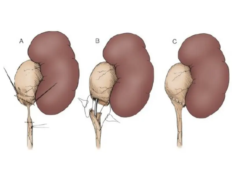
Ureteropelvic junction (UPJ) obstruction refers to a narrowing or blockage at the junction between the renal pelvis (the collecting area of the kidney) and the ureter (the tube that carries urine). The renal pelvis is located at the upper end of each ureter, which transports urine from the kidneys to the bladder. This funnel-shaped structure collects urine before it drains into the ureter. Under normal conditions, each kidney has one ureter.
What is Ureteropelvic Junction (UPJ) Obstruction?
Ureteropelvic junction (UPJ) obstruction refers to a narrowing or blockage at the junction between the renal pelvis (the collecting area of the kidney) and the ureter (the tube that carries urine). The renal pelvis is located at the upper end of each ureter, which transports urine from the kidneys to the bladder. This funnel-shaped structure collects urine before it drains into the ureter. Under normal conditions, each kidney has one ureter. The kidneys filter waste products and excess water from the blood to produce urine. Urine collects at the UPJ and is then transported through the ureters to the bladder. In UPJ obstruction, urine flow slows down or stops completely, increasing the risk of kidney damage. In most cases, only one kidney is affected.
UPJ obstruction occurs in approximately 1 out of every 1500 births and is responsible for nearly 80% of kidney swelling (hydronephrosis) caused by urinary tract obstruction. It is seen more than twice as often in males compared to females, and the left kidney is affected approximately twice as frequently as the right kidney.
Causes and Symptoms of Ureteropelvic Junction (UPJ) Obstruction
Symptoms of ureteropelvic junction obstruction include:
- Swelling on the side of the abdomen
- Febrile urinary tract infection
- Pain in the upper abdomen and back, usually after drinking fluids. The pain results from urine accumulation causing pressure on the kidney and surrounding tissues. In some cases, when the obstruction is partial, the pain may come and go as urine flow occasionally becomes possible.
- Increased risk of kidney stones
- Blood in the urine
- Nausea and/or vomiting
- Poor growth and developmental delay in children
UPJ obstruction indicates that the ureter or kidney structures did not form properly while the baby was developing in the womb. A hereditary predisposition may also play a role. UPJ obstruction may be congenital or may develop later due to causes such as infection.
In general, UPJ obstruction may result from the following causes:
- The ureteral opening is congenitally too narrow: there are abnormalities in the number or arrangement of the small muscle cells in the ureter. These cells are responsible for the muscular contractions that push urine from the kidney to the bladder.
- Abnormal folds in the ureteral wall may act like valves.
- Kinking along the ureter may develop.
- The ureter may connect to the renal pelvis at an abnormally high position, creating an abnormal angle between the kidney and ureter.
- Abnormal crossing blood vessels may compress the UPJ or disrupt urine flow.
- More rarely, in adults, UPJ obstruction may occur due to kidney stones, upper urinary tract infections, surgery, abnormal crossing blood vessels, or swelling in the urinary tract.
Diagnosis of Ureteropelvic Junction (UPJ) Obstruction
An ultrasound examination performed before birth may reveal UPJ obstruction. Urine accumulation caused by the obstruction results in abnormal enlargement of the kidney, a condition known as hydronephrosis. After birth, blood tests such as blood urea nitrogen (BUN) and creatinine levels, along with urine tests, are necessary to evaluate kidney function and urinary drainage.
Intravenous pyelography (IVP), also known as contrast kidney imaging, renal scintigraphy (nuclear renal scan), ultrasonography, computed tomography (CT), and magnetic resonance imaging (MRI) can demonstrate urinary tract obstruction and show the structure of the kidneys, ureters, and bladder.
Treatment of Ureteropelvic Junction (UPJ) Obstruction
In many infants with UPJ obstruction, the condition may resolve spontaneously within the first 18 months of life. During this period, repeated ultrasounds and scans are used to monitor the condition and ensure that permanent kidney damage does not occur. If urine flow does not improve after the first 18 months and the obstruction persists, surgery is usually required.
The most commonly performed surgery in infants is called open pyeloplasty. In this procedure, the obstructed segment of the UPJ is removed, and the ureter is reconnected to the renal pelvis with a wider opening. The goal of surgery is to allow urine to flow freely, eliminate symptoms, and reduce the risk of infection.
This operation can also be successfully performed using minimally invasive pyeloplasty, laparoscopic surgery, or robotic surgery. Another option is an endoscopic approach in which a special wire is inserted through the ureter to reach the obstructed area. The obstruction is then cut using electrical current, and a stent is placed for several weeks during healing. This procedure does not require any surgical incision, but its success rate is lower and it may need to be repeated multiple times.
Open pyeloplasty usually takes several hours to complete and has an approximately 95% success rate. Patients may need to stay in the hospital for two or three days after surgery. A drainage tube may be temporarily placed into the kidney or ureter to help urine flow during recovery. The success rates of laparoscopic and robotic surgery are similar to those of open pyeloplasty; however, these techniques require greater surgical expertise and depend on the patient’s age and the severity of the narrowing.
The information here is for general education only and is not medical advice. Please consult your physician for personalized treatment.