









Male Infertility
Infertility is defined as the inability to conceive after one year of unprotected intercourse. The frequency of infertility varies by region, but generally, infertility affects 15% of all couples. In 35% of cases, the female alone is responsible, in 30% the male alone, and in 20% both male and female factors are responsible.
What is infertility?
Infertility is defined as the inability to conceive after one year of unprotected intercourse. The frequency of infertility varies by region, but generally, infertility affects 15% of all couples. In 35% of cases, the female alone is responsible, in 30% the male alone, and in 20% both male and female factors are responsible. Low sperm count, poor semen quality, or both are present in 90% of infertile men. It is known that 23% of couples who do not conceive in the first year of unprotected intercourse achieve pregnancy in the second year, and 10% in the fourth year. Even in cases of severe low sperm count (sperm concentration below 2 million mL), the pregnancy rate within two years is 7.6%. The causes of infertility can be classified into two groups: obstructive (caused by blockage) and non-obstructive (caused by other reasons not related to blockage). In infertile men, there are abnormalities in sperm morphology, count, or transport. The evaluation of infertile males should be done using rapid, inexpensive, and non-invasive diagnostic methods; in 70% of cases, the diagnosis is made through history, physical examination, hormone analysis, and sperm analysis. More detailed, expensive, and invasive diagnostic methods should be used when necessary. Recently, the deterioration in semen quality over the years has been frequently discussed in the media. The average sperm concentration, which was 113 million in the 1940s, dropped to 66 in the 1990s and continues to decline. While researchers attribute this to environmental conditions and toxins, differences in counting methods and laboratories should also be considered.
Treatment options vary widely, ranging from medication or surgery to optimize sperm production and transport, to complex assisted reproductive techniques, depending on the underlying cause. Technological advancements have made it possible to conceive a baby from a single viable sperm and egg. While 12 months of unprotected intercourse is recommended for evaluation in newlywed couples, this period may be shorter for older couples.
Hormonal Cycle
Testicular and sexual functions are regulated by the hypothalamus-pituitary-testis loop, a closed-loop system controlled by negative feedback from the testes. The hypothalamus, the primary adaptive organ, processes signals from the central nervous system (CNS), pituitary gland, and testes, releasing gonadotropin-releasing hormone (GnRH) in bursts every 70-90 minutes throughout the day. GnRH has a half-life of 2-5 minutes.
GnRH is stimulated by the release of melatonin, a hormone secreted from the pineal gland in the brain, while male hormone, inhibin (a hormone secreted from the testes), corticotropin-releasing hormone, drugs, disease, and stress suppress GnRH secretion. GnRH enters the pituitary gland circulation, stimulating the release of gonadotropins called luteinizing hormone (LH) and follicle-stimulating hormone (FSH). The hypothalamus-pituitary-testis cycle occurs through the excitatory and inhibitory signals of these organs. GnRH release from the hypothalamus initiates the release of FSH and LH from the pituitary gland. FSH stimulates Sertoli cells to aid in sperm production, while LH stimulates the release of male hormone from Leydig cells. This cycle is suppressed by the secretion of male hormone and inhibin. FSH and LH are glycoproteins with a molecular weight of 10,000 daltons; their alpha chains are the same as those of human chorionic gonadotropin and thyroid-stimulating hormone, while their beta chains are unique to them. FSH is present in lower amounts in the blood than LH, has a longer half-life, and is not released suddenly. The sudden, evenly spaced release of GnRH stimulates the release of FSH and LH, while a continuous release of the same amount suppresses FSH and LH release.
The hypothalamus also produces prolactin, which is released from the anterior pituitary, as well as dopamine-releasing hormone (thyrotropin-releasing hormone) and vasoactive intestinal polypeptide, which suppress prolactin release. In men, increased prolactin levels can cause breast enlargement, impaired libido, erectile dysfunction, and milk production. Prolactin suppresses GnRH release from the hypothalamus and LH and FSH release from the pituitary gland. Other mediators affecting gonadotropin (LH, FSH) release are estradiol (a female hormone that is a strong inhibitor of both LH and FSH) and inhibin, secreted from Sertoli cells, which selectively suppresses FSH release.
After being released into the systemic circulation, FSH and LH exert their effects by binding to plasma membrane receptors in target cells. LH's primary function is the release of male hormones from Leydig cells, while FSH facilitates the differentiation of sperm-forming cells via Sertoli cells.
Testosterone (SDH) secretion occurs during the day, reaching its peak within a few hours of waking up. In the body, 2% of SDH is free, 44% is bound to sex hormone-binding proteins, and 54% is bound to albumin. SDH is converted to dihydrotestosterone in both local and peripheral tissues with the help of the 5-alpha reductase enzyme, and also converted to estrogen in peripheral tissues. SDH and estradiol provide feedback for the suppression of FSH and LH.
Sperm Formation Cycle (Physiology)
The testis contains Leydig and Sertoli cells and is surrounded by a structure called the tunica albuginea, which extends into the testis and forms 200-350 pyramidal regions within it. These pyramids are filled with seminiferous tubules. A testis contains 600-1200 seminiferous tubules with a total length of up to 250 meters. Between the seminiferous tubules are Leydig cells, fibroblasts, lymphatic vessels, blood vessels, and cells called macrophages. The seminiferous tubules are composed of Sertoli cells and germ cells (sperm precursor cells), and are surrounded by peritubercles and muscle cells. Sertoli cells are cylindrical in shape, with a nucleus near the base and a prominent nucleolus. They are seated at the base of the tubule, providing support to the developing sperm cell, forming a barrier between the testis and the blood, enabling spermatogenesis, and protecting germ cells from the immune system. Sertoli cells also secrete inhibin, which suppresses androgen-binding protein (EBP) in the hypothalamus and seminiferous tubules, regulating the activity of male hormones. In addition to FSH, Sertoli cell functions are also regulated by intratesticular male hormone and signals from muscle cells surrounding the tubules.
Germ cells, the precursors of sperm, develop from the swollen genitalia in the womb and migrate to the testes before the testes descend into the scrotum. In puberty, in response to FSH stimulation, germ cells develop into spermatogonium and mature into spermatozoa (mature sperm cells). The entire process from spermatogonium to spermatid takes 74 days and occurs in 14 different stages; as they mature, they move closer to the seminiferous tubule lumen and accumulate there.
Spermatogonia are located on the basement membrane of the fallopian tube wall, possess a dense nucleus and a distinct nucleolus, and come in three types: dark A, pale A, and B cells. The A cells are the mother cells; they divide to produce more dark A cells or transform into pale A cells every 16 days. Pale A cells mature into B spermatogonia. B spermatogonia undergo mitosis to develop into first spermatocytes, which are distinguishable by their large central nuclei and bead-like genetic material. Mitosis does not occur through complete separation; rather, it is a division where the intercellular bridges of the daughter cells are maintained, thus ensuring that communication is not interrupted during maturation.
The first spermatocytes undergo meiosis as they pass through the preleptotene, leptotene, zygotene, and pachytene stages, transforming into secondary spermatocytes. During this process, the cells move from the base of the fallopian tube towards the lumen in the center. The nuclei of the secondary spermatocytes are smaller. The secondary spermatocytes undergo a second meiotic division and become spermatids. Since one chromosome from each chromosome pair remains in the cell during meiosis, the chromosome pairs remain single-chromosome, and each spermatocyte develops 4 spermatid cells through one mitosis and 2 meiotic divisions.
The next step is sperm formation, continuing through stages called Sb1, Sb2, Sc, Sd1, and Sd2. Excess matter in the cell nucleus decreases, and structures called the acrosome and flagellum develop. The acrosome, like the Golgi organelle in normal cells, covers the front part of the cell nucleus and carries the enzymes necessary for penetration into the egg. Mature spermatids are close to the cavity in the center of the seminiferous tubules, and their oval-shaped nucleus stains darkly. After separating from Sertoli cells, spermatids pass through straight tubes, the rete testis, and efferent tubes to reach the epididymis.
The epididymis is 3-4 cm long, while the total length of the tubules within it reaches 4-5 meters. Sperm enter the epididymis from the head and mature as they travel towards the tail, reaching the capacity to enter the egg. The transit time through the epididymis varies between 1 and 12 days, depending on age and frequency of sexual activity. The epididymis also provides the sperm with substances such as glycerophosphorylcholine, carnitine, and sialic acid for nourishment and protection during transit. After leaving the epididymis, the sperm's journey continues in the vas deferens, which is 30-35 cm long. The function of the vas deferens is to store and transport sperm; it also secretes fluids. During sexual activity, sperm move forward peristaltically through the distal part of the vas deferens into the wider region. Here, the vas deferens merges with the seminal vesicle, a sexual gland, forming the ejaculatory duct until it opens into a bulge called the verumontanum within the prostatic urethra.
During ejaculation, the muscles surrounding the ejaculatory duct and the bulbourethral muscles adjacent to the prostate contract, and the ejaculate is expelled in a forceful manner. The contraction and closure of the bladder neck during this process is necessary for the ejaculation to be forward. The normal ejaculate volume is between 1.5 and 5 ml, with a pH level of 7.05-7.8. The seminal vesicles provide 40-80% of the ejaculate (also called semen). The secretions of the seminal vesicles in semen include fructose, prostaglandins, and other chemicals that nourish sperm, as well as bicarbonate, which buffers the acidic environment in the vagina. The fructose concentration in ejaculate is 120-450 mg/dL. Low levels indicate ejaculatory duct obstruction or congenital absence of seminal vesicles. Prostatic secretions constitute 10-30% of the ejaculate volume. It contains enzymes and proteases that dissolve clots in the ejaculate within 20-25 minutes. The prostate also secretes zinc, phospholipids, phosphatase, and spermin into the ejaculate. The portion of the ejaculate volume originating from the testes and epididymis, including sperm, is only 5%. The contribution from the bulbourethral and periurethral glands, not mentioned above, is between 2-5%, and their functions are to lubricate the urethra, buffer urine waste, and reduce acidity. Their secretion is important for sexual function.
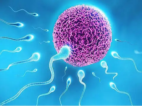
For fertilization to occur, sperm must reach the cervix, penetrate the mucus layer there, reach the uterus and fallopian tubes, enter the egg (oocyte), and dissolve the outer layer of the egg cell called the zona pellucida with enzymes present in the acrosome at the head of the sperm, and transfer the genetic material from the nucleus at the back of the acrosome into the oocyte. The stages of fertilization, the attachment of the sperm to the oocyte (female egg), and the transfer of the sperm's genetic material into the oocyte are called fertilization. Due to changes in the mucus (mucous substance) in the cervix during the female ovulation cycle, sperm entry is much easier during ovulation. After fertilization, the fertilized egg implants in the uterus. Problems at any of these stages cause infertility.
Causes and Diagnostic Methods of Infertility
Serious diseases that are more common in infertile men:Many men whose primary complaint is infertility have significant underlying health problems such as pituitary adenomas, hormone-secreting tumors, testicular cancer, liver and kidney failure, and cystic fibrosis. It is necessary to determine the presence of these conditions during the evaluation of these patients. Additionally, the incidence of cancer is increased in infertile men. Overall, 2.2% of azoospermic men and 1.1% of non-azoospermic infertile men developed cancer during a 7-year follow-up period. The incidence of cancer in non-azoospermic infertile men was close to or slightly increased compared to the normal population. The highest incidence of cancer was observed in azoospermic men. The cancers that developed included prostate, testicular, central nervous system, skin, and stomach cancers.
Gender:Female-only infertility accounts for 35% of infertility cases, while male-only infertility accounts for 30%. Problems in both partners occur in 20% of cases, and the cause remains unknown in 15%. Even if a very clear cause of infertility is found in one partner, the other partner must also be evaluated. Additionally, the sexual function of both partners should be assessed.
Age:The effects of age on reproduction are not definitively known. As a man ages, levels of male hormones (STEM) decrease, while levels of estradiol (female hormone) increase. Studies show that sperm density decreases with age. While spermatids are found in 90% of the seminiferous tubules of young men, this number drops to 50% between the ages of 50-70, and to 10% at age 80. Furthermore, around age 50, 50% of Sertoli cells are lost, and at 60 years of age, the number of Leydig cells decreases by 50%. Despite this, the frequency of pregnancy in older men is similar to that in younger men, but the time to conception may be longer.
1-Story-Resume-Inquiry
A complete medical and urological history should be conducted when evaluating infertile men. Points to highlight include the duration of infertility and previous reproductive history. Questions should be asked about the couple's sexual habits, their knowledge of the optimal time for pregnancy, and their use of medications and lubricants that may have harmful effects on sperm. Male patients should be asked about testicular torsion, a history of childhood puberty, developmental delay, precocious puberty, urinary infections, sexually transmitted diseases, and bladder neck surgery. A history of neurological diseases, diabetes, and lung infections should also be obtained. Loss of sense of smell, milk production from the breasts, visual field defects, and sudden loss of sexual appetite may be signs of a pituitary tumor.
Adolescence (early, normal, or delayed):In boys, puberty occurring before the age of 9 is considered precocious puberty and may be indicative of significant hormonal disorders. Precocious puberty may be a sign of hormonally active tumors of the testes, adrenal glands, or pituitary gland, or of adrenal gland enlargement. Conversely, delayed puberty may indicate insufficient production of male hormones due to hypothalamic, pituitary, or testicular failure, or unresponsiveness to male hormones in the target organ.
Childhood illnesses or past surgeries:Unilateral or bilateral undescended testicles impair sperm production and semen quality, regardless of the timing of the operation. If the patient has hypospadias, sperm may not reach the cervix; exposure to diethylstilbestrol (a former birth control pill) in utero can cause epididymal cysts and undescended testicles; previous bladder neck surgeries can cause ejaculate to move back into the bladder during ejaculation; testicular veins or vas deferens may have been damaged during inguinal hernia, hydrocele, or varicocele surgeries; testicular torsion and trauma can cause testicular shrinkage and the formation of antisperm antibodies. All these conditions should be thoroughly investigated.
Medical historyIn men, poor general health also affects reproductive health. Diseases that affect reproductive ability include:
- Diabetes mellitus can cause autonomic neuropathy, neurogenic erectile dysfunction, and retrograde ejaculation (semen not exiting the urethra during ejaculation, flowing back towards the bladder).
- Obesity (excessive weight gain) disrupts hormonal metabolism, causing excessive conversion of the male hormone estrogen to estrogen in peripheral tissues. Increased estrogen reduces the intensity of the rhythmic release of LH hormone, leading to a decrease in sperm density.
- In sickle cell anemia, testicular damage can occur due to anemia. In thalassemia (Mediterranean anemia) and sickle cell anemia, repeated blood transfusions can cause hemosiderosis, a condition characterized by iron retention in tissues, which can lead to infertility.
- Chronic kidney failure can cause hypogonadism (underactive testes in men) and changes in physical appearance, such as feminine features (hair loss, breast enlargement, and a higher-pitched voice).
- Liver diseases can impair secondary male characteristics and, with increased estrogen levels, can cause testicular shrinkage and breast enlargement.
- Hemochromatosis can be a cause of hypogonadysfunction, leading to a deficiency in androgen hormones and an increase in estrogen levels.
- If mumps develops after puberty and causes testicular involvement, it can shrink the testicles and lead to infertility.
- Sexually transmitted diseases and tuberculosis can cause obstruction in the vas deferens and epididymis.
- Mycoplasmas are sexually transmitted infections that bind directly to sperm, reducing sperm motility.
- Smallpox (no longer seen), inflammation of the prostate, testicles, seminal vesicles, or urethra can cause obstructive azoospermia.
- Acute or chronic medical illnesses
- Patients with infertility should be asked if they have had a febrile infection, as sudden febrile illnesses reduce FSH and LH secretion. This, in turn, impairs sperm production for 1-3 months.
- Anesthesia, surgery, starvation, heart attack, hepatic coma, head trauma, stroke, respiratory failure, heart failure, sepsis, and burns likely increase the release of dopamine and opioid substances, thereby suppressing the release of gonadotropins.
- Chronic medical conditions directly suppress sex hormone production and sperm production, leading to organ failure.
Sexual history:Couples should be questioned about the timing, frequency, and method of intercourse, as well as their knowledge of the woman's ovulation cycle. Studies have shown that the most favorable time for pregnancy is intercourse every 48 hours in the middle of the ovulation period. Lubricants such as KY gel and saliva are toxic to sperm. While egg white, hazelnut oil, vegetable oils, and petroleum-derived gels are not considered toxic to sperm, they should be used in small quantities if necessary.
Testicular cancer:Testicular cancer can cause infertility due to the effects of the tumor on the affected area or as a result of testicular removal. In 60% of patients diagnosed with testicular cancer, a reduced sperm count is found. Germ cell tumors (the cells that produce sperm), congenital absence of a testicle, androgen deficiency, and undescended testicles all impair testicular function. In patients with testicular cancer, sperm production disorders are also seen in the healthy testicle. Even after surgical removal of the cancerous side, sperm production disorders in the healthy testicle often persist.
Testicular cancer treatment:High-dose chemotherapy affects germ cells. Alkylating chemotherapy drugs such as cyclophosphamide, mustin, and chlorambusil cause severe damage to the seminiferous tubules and destroy sperm stem cells. It should also be kept in mind that chemotherapy causes mutations in chromosomes; therefore, sperm preservation before chemotherapy is recommended for such patients, or pregnancy attempts are advised to wait at least one year after the end of treatment. Retroperitoneal lymph node dissection (RPLND); RPLND, applied to some patients with testicular tumors, causes ejaculation disorders and/or retrograde ejaculation. Radiation therapy affects type B spermatogonia and possibly spermatocytes. Even low radiation doses such as 0.15 Gy can cause irreparable testicular damage, but complete recovery may be possible if not all stem cells are destroyed. While sperm production may return 9-18 months after radiotherapy below 1 Gy, this period can extend to 4-6 years at doses of 5 Gy. In two-thirds of patients, fatherhood can occur even with radiotherapy and chemotherapy if ejaculation function is intact. In first-degree germ cell testicular tumors, only the affected testicle is surgically removed to minimize treatment-related side effects, and the patient is monitored and regularly checked. In more advanced stages, the risk of retrograde ejaculation increases due to RPLND (Retinopathy of Prematurity). After surgical removal of the testicle, improvement in semen parameters begins. This effect is thought to be due to the patient's relief from anxiety and the elimination of the harmful effects of tumor-secreted substances on sperm production. In patients with a single testicle, if there is a tumor in that single testicle, partial surgery can be performed leaving the tumor-free portion intact, thus preserving fertility. Healthy, tumor-free tissue removed after surgery can be separated and frozen for future use in assisted reproductive techniques (ICSI, IVF).
Social history:Smoking and marijuana use reduce sperm density, motility, and morphology. The use of anabolic steroids for muscle building causes hypogonadism and is associated with structural and genetic sperm damage. Alcohol causes a sudden and chronic decrease in male hormone production. Emotional stress interrupts GnRH release and causes hypogonadism. Exposure to high heat in saunas, Turkish baths, or while working causes temporary reductions in sperm production. Contrary to popular belief, tight underwear does not reduce fertility. Although wearing tight underwear causes a 0.8-1 degree Celsius increase in testicular temperature, sperm parameters do not change, and sperm production is not impaired.
Medicines:Drugs that impair reproduction (fertility) include: Spironolactone, cyproterone, ketoconazole, and cimetidine, which have antiandrogenic effects. Tetracycline reduces male hormone levels by 20%. Nitrofurantoin suppresses spermatogenesis (sperm production). Sulfasalazine temporarily reduces sperm motility and density. Colchicine, methadone, methotrexate, phenytoin, thioridazine, and calcium channel blockers can cause infertility.
Family History:Congenital diseases should be considered if there is a family history of midline defects, undescended testicles, hypogonadism, and testicular atrophy. A family history of cystic fibrosis or hypogonadism should be investigated in infertile men.
Respiratory Tract Diseases:In the presence of infertility and recurrent respiratory tract infections, immotile cilia syndrome or Kartafener syndrome, characterized by sperm immobility, should be considered. Cystic fibrosis is one of the causes of congenital obstructive azoospermia due to the absence of both vas deferens. If there is a deficiency in both parts of the recessive CFRD gene inherited from the mother and father, cystic fibrosis is clinically observed; if there is a deficiency in only one parent, bilateral congenital absence of the vas deferens may occur even if cystic fibrosis is not clinically observed. In Young's syndrome, lung infections are frequent due to thickened secretions, and obstruction develops in the epididymis, resulting in obstructive azoospermia.
Environmental and Occupational Exposure:Many rodenticides cause estrogen-like effects. Dibromochloropropane, frequently used against parasites in agricultural production, can cause irreversible azoospermia through unknown mechanisms. Lead suppresses hypothalamus-pituitary communication. Carbon disulfide, used in the rayon industry, causes changes in semen (ejaculate), hypothalamus, and pituitary gland. Workers in the steel and ceramics industries experience decreased spermatocyte maturation due to high temperatures.
Spinal Cord Injury:Severe spinal cord injuries can cause anejaculation (inability to ejaculate). In these men, electroejaculation or sperm retrieval methods are used in treatment. Semen quality also progressively deteriorates in such patients; one year after spinal cord injury, semen in most patients consists of immobile sperm, due to the influx of inflammatory cells into the semen. While 26% of sperm in the ejaculates of spinal cord injured patients are found to be viable and only 14% motile, seminal samples taken from the seminal vesicles with a needle show 74% viability and 54% motility. The reason for this is the lack of stimulation of the prostate by the nervous system in these patients and the overstimulation of the immune system due to non-infectious causes. Infertile patients with spinal cord injuries have also been found to have abnormalities in sperm nucleus maturation and DNA integrity.
2-Physical Examination
Testes:The length and consistency of one testicle are evaluated and compared to the other. An approximate testicular volume is estimated; the value should be greater than 20 ml. The length should be at least 31 mm. Testicular shrinkage may be a sign of primary testicular failure, Klinefelter syndrome, hormonal disorders, post-pubertal mumps, liver disease, and muscular dystrophy. Swelling accompanied by pain may be a sign of orchitis (testicular inflammation), while painless swelling may be a sign of a testicular tumor, testicular tuberculosis, or end-stage syphilis.
Epidemic:The presence of growths and cysts in the epididymis may indicate a blockage in the sperm ducts, while pain may be a sign of an infection in the epididymis.
Vas deferens:Both sides should be examined manually along their entire length to check for any absence, partial swelling, thinning, or other changes. Bilateral congenital absence is seen in cases of deficiency in one or two copies of the cystic fibrosis gene, while partial absence of the vas deferens may be seen in cases of mutations in the cystic fibrosis gene. It may be seen along its entire length with hard nodules due to tuberculosis involvement, and the presence of a previous vasectomy is evident during examination.
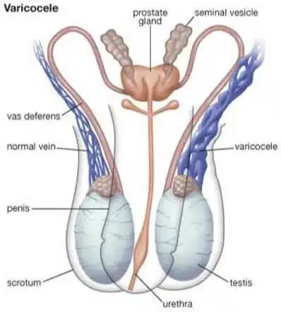
Spermatic cord:During examination, patients should be checked for varicocele, the most common cause of infertility that can be corrected surgically. Varicocele is assessed by maneuvers that increase intra-abdominal pressure while standing. A varicocele located on the right side, or remaining stable without increasing with increased intra-abdominal pressure, may indicate intra-abdominal tumors or vascular occlusions.
Penis:During a penile examination, the presence of narrowing at the urethral opening, and congenital anomalies called hypospadias and epispadias are noted, as these conditions can cause problems with sperm transfer to the vagina. Any curvature of the penis and hard plaques are also recorded. Hypospadias, which prevents sperm cells from collecting at the cervix, can be a cause of infertility.
Rectal Examination:The prostate is examined for enlargement, hardness, and any masses. If the ejaculatory ducts are blocked, the seminal vesicles may be palpable.
Body shape:In patients with hormonal imbalances, a condition known as eunuchoid appearance is characterized by reduced hair growth, weak muscle structure, and a long, thin body structure, particularly in the lower extremities, due to delayed epiphyseal closure. Obesity, particularly noticeable around the navel, stretch marks on the abdominal skin, and a moon face appearance suggest Cushing's syndrome, an adrenal gland disorder. Breast enlargement, milk production, headaches, and visual field defects suggest pituitary tumors. A neck examination may reveal an enlarged thyroid gland and palpable murmurs in the neck vessels. Liver enlargement and enlarged lymph nodes, if present, can also be detected by palpation.
3. Causes of Infertility
a- Causes of pretesticular infertility:
Pretesticular causes of infertility are congenital diseases of the hypothalamus and pituitary gland, or conditions causing infertility due to organ diseases that disrupt the hypothalamus-pituitary axis. Hypothalamic diseases cause hypogonadotropic hypogonadism. If GnRH is not secreted, LH and FSH hormones are not secreted from the pituitary gland. These patients respond to exogenously administered GnRH and human chorionic gonadotropin (hCG), an LH-like hormone.
Idiopathic (of unknown cause) hypogonadotropic hypogonadism:In the absence of other disorders, unexplained deficiency in GnRH secretion or loss of smell, midline defects such as cleft lip and palate, hearing impairment, undescended testicles, and color blindness can be seen as part of Kalmann syndrome. Kalmann syndrome can be familial (X-linked and autosomal) or occur individually. It occurs in approximately 1 in 10,000 to 60,000 births. It is thought to be caused by the failure of GnRH-secreting nerve cells to migrate to their proper location in the hypothalamus. Due to delayed closure of the epiphyseal plates, patients generally have long arms and legs, delayed puberty, and small testicles. Although patients may reach normal height with male hormone therapy, sperm production does not improve. External administration of male hormones to increase sperm production should be avoided because it suppresses GnRH secretion and reduces intratesticular male hormone levels. In approximately 20% of patients, a series of sudden, frequent doses of GnRH and hCG may restore normal sperm production. In most patients, sperm production can be achieved when human FSH is added to hCG in treatment. In cases of adult-onset idiopathic hypogonadotropic hypogonadism, a response to clomiphene citrate treatment may be obtained.
Prader-Will syndrome:Patients typically present with obesity, mental retardation, small hands and feet, and hypogonadotropic hypogonadism due to GnRH deficiency. It is caused by inherited breaks in regions 11-13 (represented as 15q11-13) on the long arm of chromosome 15.
Laurence-Moon-Biedl sendromu:Patients exhibit retinal disorders and multiple fingers, and infertility occurs due to hypogonadotropic hypogonadism.
Other situations:Central nervous system tumors, temporal lobe epilepsies, and numerous medications, such as dopamine antagonist drugs, can disrupt the hypothalamic-pituitary axis at the hypothalamus level. Both hypothyroidism (underactivity) and hyperopia (overactivity) of the pituitary gland cause infertility. Hypothyroidism (underactivity) can be congenital or acquired. Conditions that cause acquired pituitary insufficiency include tumors, obstruction of pituitary vessels, radiation, and infection. Non-secretory pituitary tumors can cause pituitary insufficiency by compressing the gland, while secretory pituitary tumors can hinder the normal functioning of the pituitary gland through irregular gonadotropin secretion or excessive prolactin secretion.
Prolactinoma:Prolactin-secreting adenomas are the most common pituitary tumors that secrete prolactin. Prolactin stimulates breast development and milk secretion; therefore, male children with prolactinomas experience breast enlargement and milk secretion. Bilateral peripheral vision loss should suggest an enlarged pituitary tumor compressing the optic nerve crossing. Prolactin levels higher than 150 microg/L suggest a prolactinoma, while prolactin levels higher than 300 microg/L almost immediately confirm the diagnosis. CT or MRI scans of the pituitary gland should be performed to determine the size of the tumor.
In patients with small adenomas, drugs with dopamine-like effects, such as bromocriptine and cabergoline, are used to lower prolactin levels. In some patients, male hormone levels increase, and sperm counts normalize. Surgery for small tumors is successful in 80-90% of cases, but adenomas recur in 17% of patients. Surgical treatment of large prolactinomas is rarely curative; therefore, it should be recommended in patients with significant visual field deficit and those who cannot tolerate bromocriptine treatment.
Isolated LH deficiency (fertile enucoid):In these patients, FSH levels are normal while LH levels are low; they have an enuccoid appearance (long, thin, hairless, and weakly muscular); testicular size is normal, but ejaculate volume is low. They are treated with external hCG administration.
Isolated FSH deficiency:It is a very rare cause of infertility. Patients have oligospermia (sperm concentration less than 2 million/ml) along with normal LH levels and low FSH levels. Treatment involves administering human menopausal gonadotropin (hMG) or FSH.
Thalassemia (Mediterranean anemia):Thalassemia patients frequently receive blood transfusions due to impaired red blood cell production. During these transfusions, excessive amounts of iron enter the body, accumulating in the pituitary gland and testes, causing tissue damage and leading to both pituitary and testicular failure. Treatment involves gonadotropins and iron-binding therapy.
Cushing's disease:The suppressive effect of excessive cortisol secretion from the adrenal gland reduces GnRH secretion from the hypothalamus.
Peripheral organ diseases:The hypothalamus-pituitary axis can be disrupted by hormone-secreting peripheral tissue tumors, or by underactive or overactive adrenal glands, or by excessive estrogen levels. Causes of excessive cortisol production include adrenal gland hypertrophy, adenomas (benign hormone-secreting tumors), cancers, or lung cancer. Excessive steroid use due to ulcerative colitis, asthma, arthritis, or organ transplants can also lead to elevated cortisol levels.
Adrenal cortex deficiency can develop due to infection, circulatory disorders, or congenital enlargement of the adrenal cortex. Congenital deficiencies in several enzymes involved in hormone production in the adrenal gland, most commonly the 21-hydroxylase enzyme, also lead to adrenal enlargement and dysfunction. When cortisol secretion from the adrenal gland and its suppressive effect on the pituitary gland are absent, adrenocorticotropic hormone (ACTH), secreted from the pituitary gland to stimulate cortisol production in the adrenal gland, is excessively secreted. This excessive hormone secretion from the adrenal gland suppresses GnRH secretion from the hypothalamus. Individuals with 21-hydroxylase deficiency have a short neck, early puberty, and small testes. Blood levels of 17-hydroxylase and urinary excretion of 17-ketosteroids are increased in these patients.
Sertoli and Leydig cell tumors, liver failure, and obesity are causes of excessive estrogen secretion. High estrogen suppresses the pituitary gland, reducing LH and FSH secretion.
b-Testicular Causes:
Testicular causes include those related to chromosomal changes or non-chromosomal causes. Chromosomal causes are generally associated with anomalies of the sex chromosomes.
Chromosomal abnormalities:While the prevalence of chromosomal abnormalities in the general population is 0.6%, it is 6-13% in infertile men. This rate increases to 10-15% in men with azoospermia or severe oligospermia. In men with non-obstructive azoospermia or severe oligospermia, karyotype testing (46XY) and genetic evaluations to determine the presence of Y chromosome microdeletions should be performed.
-Klinefelter syndrome:Klinefelter syndrome is the most common chromosomal anomaly causing male infertility, occurring in 1 in every 500-1000 male births. Patients have an extra X chromosome, resulting in a karyotype of 47,XXY. The cause is usually the failure of chromosome separation during meiosis between the mother and the baby. Currently, the only known risk factor for Klinefelter syndrome is maternal age. The karyotype analysis in Klinefelter syndrome shows 47,XXY chromosomes, indicating an extra X chromosome instead of the required 46. Infertility in Klinefelter syndrome is primarily due to testicular failure, and most patients are azoospermic. Hormone tests show increased gonadotropin levels, while 60% of cases have low blood testosterone levels. Interestingly, libido (sexual appetite), erection, and orgasm are normal in most patients. Therefore, treatment with male hormones has little effect. If external male hormones are administered, it can further reduce sperm production. Physical examination reveals breast enlargement (gynecomastia), small testes, enucoid body structure, and delayed puberty. Although some patients have secondary sexual characteristics such as hirsutism, muscle development, and deepening of the voice, their history shows late onset and late completion of puberty. The frequency of breast cancer, leukemia, diabetes, pituitary diseases, and pituitary tumors is increased in these patients. Biopsies show loss of the tubular structure in the seminiferous tubes. In some men with Klinefelter syndrome, pregnancy may be possible with assisted reproductive techniques. In 20% of azoospermic men with Klinefelter syndrome, sperm production foci may be found in the testes on biopsy. Although 47, XXY is seen in spermatogonia and primary spermatocytes, normal chromosomal structure is seen in secondary spermatocytes and spermatids. In these patients, genetic testing can be performed on embryos created with assisted reproductive techniques before implantation in the mother's uterus.
-XX male (reverse gender syndrome):In these patients, the condition occurs due to the crossover of the SRY gene, which determines sex, from the Y chromosome to the other X chromosome or to a non-sex chromosome. Patients are short in stature, have small, firm testes, and breast enlargement; their penis length is normal. Azoospermia is present, and if the testes have not descended into the scrotum, the risk of developing testicular cancer is high. Scarring of the seminiferous tubules is observed. The SRY gene, located on the short arm of the Y chromosome, determines sex. Mutations in the SRY gene region or its transfer to other chromosomes are the cause of sex abnormalities and infertility.
-XYY male:It occurs in 0.1-0.4% of newborn males. These patients are often tall and severely oligospermic or azoospermic. Biopsy reveals arrest in the maturation stages of sperm development or the absence of germ cells. If present, the karyotype of the sperm cells may be normal.
-Noonan syndrome (46, XY):This syndrome affects patients with a 46,XY karyotype but is caused by mutations in only four genes. Mutations in the PTPN11, SOS1, RADF1, and KRAS genes result in a broad neck, short stature, lowered ears, drooping eyelids, a broad chest, lymphedema in the hands and feet, cardiovascular anomalies, and cubitus valgus (hand anomaly). Leydig cell function is impaired, and most patients are infertile due to primary testicular failure.
-Mixed gonadal dysgenesis (mixed gonadal developmental disorder, 45, X/ 46, XY):Patients often exhibit genitalia of both sexes, with a testis on one side and a thinned gonad in the form of a line on the opposite side.
-Androgen receptor dysfunction:Androgen receptors (structures or receptors to which male hormones bind and exert their effects) are essential for sperm production; therefore, dysfunction of these receptors leads to infertility. In Reifenstein syndrome in men, partial unresponsiveness of androgen receptors is observed along with a wide spectrum of anomalies in the external genitalia and infertility. Sperm production is impaired because the sperm-producing cells do not respond sufficiently to male hormones. Since this does not exert the same pressure on the hypothalamus-pituitary axis as a normal testis, gonadotropin (FSH and LH) and male hormone levels are elevated. It is thought that these receptor dysfunctions result from deficiencies in specific regions of certain chromosomes.
-Y chromosome microdeletion syndrome:The long arm of the Y chromosome, particularly the Yq11.23 region, is thought to be closely associated with reproductive capacity. While it is known that large deletions in the long arm of the Y chromosome cause azoospermia, it has recently been found that small deletions of various types are also a significant cause of infertility. These deletions cannot be detected in genetic tests where normal karyotype analysis is performed (karyotype analysis only evaluates the total number of chromosomes and which sex chromosomes are present). This is because polymerase chain reaction (PCR) base-sequence labeling studies or southern term blot tests are required to detect chromosomal deletions. Three regions called azoospermia factor a, b, and c have been identified on the long arm of the Y chromosome (AZFa, AZFb, AZFc). These deletions can be demonstrated in 3-19% of patients with idiopathic (unexplained) infertility and 6-14% of patients with oligospermia, while in patients diagnosed with infertility due to other causes, deletions can be shown in approximately 7% of cases. These tests should be performed on patients with azoospermia and severe oligospermia who are planning to undergo assisted reproductive techniques.
-Bilateral testicular absence (vanishing testes):Males are born with a normal 46, XY karyotype and the absence of both testes. Their male-type appearance indicates the presence of male hormones in utero. While the cause is unknown, possible causes include infection, vascular diseases, or bilateral testicular torsion (a condition where the testes twist several times around themselves, disrupting blood flow and causing testicular shrinkage). Karyotype analysis shows the presence of the normal SRY gene (sex-determining region on chromosome 8). Although externally administered male hormones can achieve normal male development, infertility does not resolve.
- Down Syndrome:These patients have moderate testicular failure along with a decrease in germ cells (sperm precursor cells). LH and FSH levels are generally found to be elevated.
-Myotonic dystrophy:This condition, characterized by delayed relaxation of muscles after contraction, is caused by a deficiency in the dystrophin gene, which is autosomal dominant (non-sex-linked). Due to damage in the seminiferous tubules, testicular atrophy and primary failure are present in 75% of patients. Scarring of the seminiferous tubules is seen on biopsy. Currently, there is no effective treatment.
Non-genetic causes of testicular infertility:Non-genetic causes of testicular failure can be idiopathic or caused by factors that damage the testicle, such as medications, radiation, orchitis (inflammation of the testicles), trauma, or torsion.
- Varicocele:It is caused by dilation of the vascular structures called pampiniform plexuses within the scrotum. It is seen in 15% of men. It is the most treatable cause of infertility (30-35%). It is the most frequent cause of acquired infertility (75-85%). It is generally seen on the left side; if it is only seen on the right side, intra-abdominal masses should be considered. It usually does not cause symptoms; in most men, testicular atrophy and infertility are not present at the time of diagnosis. However, it is thought that sperm production and hormone production are impaired due to increased intratesticular temperature, accumulation of toxic substances in the testicles, and/or reduced oxygen levels in germ cells, and this gradually increases over time. In addition, since the effect of insulin-like growth factor (IGF) on semen quality has been demonstrated, its role in varicocele is being investigated. In one study, it was observed that IGF increased after varicocelectomy surgery, reaching the level of normal fertile men; therefore, it is thought that IGF plays a role in varicocele-related infertility. In varicoceles, the increased frequency of immature sperm, apoptosis, and dead sperm indicates severe abnormalities during meiosis, and these abnormalities generally resolve after surgery. Following varicocelectomy, regardless of the severity of infertility, semen parameters improve in 40-70% of patients, and pregnancy occurs in 40% of patients without the need for further treatment. In newly pubertal men, surgery should be recommended when varicocele is detected. However, there is no consensus on whether this surgery should be performed only when testicular shrinkage occurs or as a routine procedure. In patients with azoospermia, sperm is found in the semen of 33% after varicocelectomy, but azoospermia recurs within a few months; therefore, in such patients, semen cryopreservation should be performed when sperm is found in the semen. For more information, click here: https://www.hakkiperk.com/tr/hizmet/detay/163/varikosel
-Undescended/Ectopic testis:Undescended testicles, found in 3% of full-term male babies, decrease to 1% by the end of the first year. Undescended testicles can occur alone or be a symptom of another syndrome such as Prune Belly syndrome. Even if the testicle is surgically brought down, the frequency of infertility is higher than normal in these patients because there is a possibility of a hereditary defect in the testicle itself. The higher the testicle is located and the longer it remains outside the scrotum, the higher the risk of infertility. Biopsies show a decreased number of Leydig cells in the testicle, along with a decrease in sperm production. Even if undescended testicles are unilateral, the congenital defect can be present in both testicles; therefore, sperm counts in these patients are generally lower than expected. For more information, click here: https://www.hakkiperk.com/tr/hizmet/detay/198/%C4%B0nmemi%C5%9F-Testis
-Trauma:This is the most common acquired cause of infertility. It can also be physical, due to trauma, heat trauma, or separation from the body.
-Sertoli Cell Only Syndrome (or absence of germinal cells):In the absence of germinal (sperm precursor) cells, LH and testosterone levels are normal while FSH levels are elevated. The exact cause is unknown, but it is thought to be multifactorial. Patients have small to normal testicular size and azoospermia. Secondary sexual characteristics (muscle structure, hair growth, beard, and voice deepening) are normal. Testicular biopsies show that the seminiferous tubules are lined only with Sertoli cells; germ cells are not seen.
-Chemotherapy:Chemotherapy is toxic to actively dividing cells. This is because chemotherapy drugs target rapidly dividing cells, as cancer cells divide quickly. Testicular germ cells are also rapidly dividing cells and are therefore affected by chemotherapy drugs. The chemotherapy drugs that most commonly cause infertility are those in the alkylating agent group, such as cyclophosphamide. For example, in Hodgkin's lymphoma, the probability of infertility with chemotherapy ranges from 80-100%.
-Radiation therapy:Leydig cells divide less frequently than germ cells and Sertoli cells, so they are not significantly affected by radiation therapy. Sertoli and germ cells, however, are highly sensitive to radiation. If the progenitor cells survive radiotherapy, sperm production may begin a few years later. However, due to the possibility of radiotherapy causing chromosomal abnormalities, patients should be advised to avoid pregnancy for up to two years. Even if the testes are protected with lead plates during radiotherapy, post-radiation infertility can occur due to free oxygen radicals.
- Orchitis (testicular infection):One of the most common causes of acquired testicular failure is viral orchitis in adulthood, such as mumps, echovirus, or group B arbovirus. Mumps orchitis develops in 25% of adults who contract mumps, with bilateral involvement in one-third and unilateral involvement in two-thirds. In mumps, orchitis can begin a few days after infection of the parotid gland, or the infection can start in the testis before the parotid gland. The virus can directly damage the seminiferous tubules, or it can cause excessive edema, leading to compression of the testicular tissue within the surrounding, rigid, and inflexible tissue, and the resulting circulatory disturbance can damage the testis. After the recovery period, the testis may return to normal or shrink. The degree of shrinkage is independent of the severity of the infection and is seen within 1-6 months. While fertility is preserved in 75% of patients with unilateral mumps orchitis, the fertility rate drops to 33% in bilateral mumps orchitis.
-Human beta-defensin disorders:Beta-defensin, a protein secreted from the epididymis, plays a crucial role in sperm maturation, and its deficiency reduces the ability of sperm to penetrate the wall of the female egg. A specific subtype, human beta-defensin 1 (HBD1), is found in all epithelial cells of the body and has preventative functions against viruses, bacteria, and fungi. HBD1 is present in seminal fluid and semen, particularly in sperm cells, especially in the lower parts of the head and the midsection. HBD1 production is reduced in patients with decreased sperm motility and those with abundant leukocytes in the semen. One study showed that in patients with low sperm motility and leukocytes in the semen who received recombinant HBD1 externally, post-treatment antibacterial activity increased, semen quality improved, and the importance of human beta-defensin in infertility treatment was highlighted.
-Other reasons:Other causes of testicular failure include testicular involvement in granulomatous diseases, such as leprosy and sarcoidosis. Sickle cell anemia causes sickling and circulatory disorders within the testicles. Excessive use of alcohol, cigarettes, caffeine, and marijuana can cause testicular failure. Despite all these factors, the cause remains undetermined in 25% of infertile men.
c- Post-testicular causes:
Post-testicular causes of infertility involve abnormalities in the transport of sperm through the fallopian tubes, which can be congenital or acquired. Blockages in the sperm transport pathways occur in 7% of infertile patients and are among the treatable causes of infertility. Other post-testicular causes of infertility include the inability of sperm to pass through the cervical mucus or structural abnormalities.
Congenital obstruction in the fallopian tube system:Maternal exposure to diethylstilbestrol during pregnancy has been shown to cause obstructions in the sperm transmission pathways in children. Regional vas deferens developmental delay is defined as the presence of at least two underdeveloped regions along the vas deferens.
Cystic fibrosis:It is the most common genetic disorder in the white race. Almost all patients with cystic fibrosis have bilateral congenital absence of the vas deferens. Other congenital anomalies of the urinary system are also common in these patients. These patients are candidates for assisted reproductive techniques after genetic screening tests.
Acquired obstruction in the tubal system:The tubes necessary for sperm transport can become blocked due to infections such as chlamydia, gonorrhea, tuberculosis, and smallpox. Young's syndrome is a disease in which blockage occurs due to excessive viscosity of secretions in the epididymis. Trauma, previous sperm retrieval methods, and surgical procedures in the groin area can lead to blockages in the sperm-carrying ducts. Ejaculatory ducts can be blocked by small stones, or sperm ducts can be blocked by pressure from prostate cysts. Vasectomy, hydrocelectomy (5-6%), and spermatocelectomy (up to 17%) operations can cause damage and blockage in the epididymis.
Antisperm antibodies:Antisperm antibodies bind to sperm, disrupting their motility and causing them to clump together. As a result, sperm cells unable to progress through the female reproductive tract cannot reach the female egg.
Whip disorders:Immotile cilia syndrome, a condition characterized by immobility of the flagellum responsible for sperm motility, can occur alone or in conjunction with Kartagener syndrome, which involves the reversal of organ placement. In respiratory system cells, the cilia and sperm flagella, which should be active, remain immobile, leading to infertility, sinusitis, bronchiectasis, and respiratory system infections.
Ejaculatory duct obstruction:Partial or complete ejaculatory duct obstruction is responsible for 1-5% of male infertility cases. Patients have palpable bilateral vas deferens on examination, but ejaculatory volume is low, and they may complain of painful ejaculation. Causes include cysts in the midline or near the prostate, calcification or stones at the ejaculatory duct opening, infections, and surgical interventions. Transrectal ultrasonography may show enlarged seminal vesicles, although not in every case. Microscopic examination of fluid obtained from the seminal vesicle under TRUS guidance to detect sperm, or radiography, is necessary for diagnosis. TRUS in an infertile azoospermic patient shows a central cyst within the prostate and enlarged seminal vesicles.
Ejaculation disorders:Absence of ejaculation (anejaculation) or retrograde ejaculation (ejaculate flowing backward into the bladder) is caused by factors such as an open bladder neck (e.g., previous prostate surgery) and the inability of the bladder neck to close during ejaculation. Causes include: diabetic neuropathy, bladder neck surgery, retroperitoneal lymph node surgery, transurethral resection of the prostate (TUR-P), colon and rectal surgery, multiple sclerosis, spinal cord injuries, and alpha-antagonist medications that cause bladder neck dilation. Absence of ejaculation or retrograde ejaculation should be considered in the following situations: a history of medication use or previous surgical procedures, low ejaculate volume, and the presence of 10-15 sperm per microscopic field in a urine sample taken after ejaculation under high magnification.
4- Laboratory Tests in Male Infertility
a-Semen analysis (spermogram):
Semen analysis is an indispensable test in the evaluation of male infertility. Abstinence from sexual activity for 2-3 days before the test improves the quality of the sample. Each day of abstinence increases semen volume by 0.4 ml, while a 7-day abstinence period can lead to a 10-15 million sperm/ml increase in sperm concentration. The semen sample should be analyzed within one hour, and 2-3 analyses should be performed at least every 2-3 days. Numerous parameters should be analyzed, including semen volume, sperm density, quality, motility, and the percentage of normal-looking sperm. The test alone does not indicate fertility. The World Health Organization (WHO) established lower and upper limits of normal semen test results in 2010. The lower limit of normal corresponds to the 5th centile, but this does not constitute a cutoff point for determining fertility/infertility.
Volume:Normal ejaculate volume is between 1.5 and 5 ml. According to the WHO, the lower limit of normal is 1.5 ml. Semen volume decreases in patients with retrograde ejaculation, congenital absence of the vas deferens or seminal vesicles, obstruction of the sperm conduction ducts, hypogonadism, or neurological conditions with poor sympathetic response. Excess volume is often due to the mixing of urine with the sample.
Semen quality:Semen, initially clotted, liquefies within 5-25 minutes with the help of enzymes secreted by the prostate and added to the semen. Semen can be dripped during this process. If the initial semen is clotted, it should suggest an ejaculatory duct obstruction or congenital absence of seminal vesicles. Semen that does not liquefy may be due to sexual intercourse prior to the test.
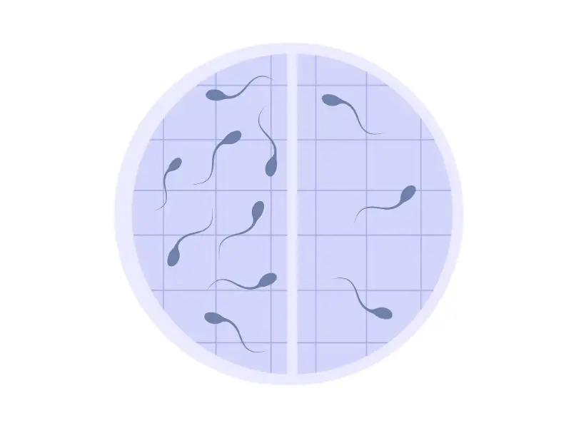
Sperm density:Normal sperm density (concentration) should be greater than 20 million per ml. The WHO's 2010 lower limit for sperm density (5th percentile) is 15 million per ml, with a total sperm count greater than 50-60 million. A sperm density of less than 20 million per ml is called oligospermia, and a density of less than 5 million per ml is called severe oligospermia. Azoospermia is the complete absence of sperm in the semen. To confirm the diagnosis of azoospermia, the entire semen should be centrifuged and examined under a light microscope for the presence of sperm. Patients with azoospermia should undergo a post-ejaculation urine test to assess the presence of sperm in their urine.
Sperm motility:This is the microscopic evaluation of the percentage of sperm exhibiting whip-like motility. Normal motility is defined as 60% of sperm moving normally. According to the WHO, the lower limit, the 5th percentile, is 40%. Grade 0 is immobile, grade 1 is sluggish motility, grade 2 is slow and non-linear motility, grade 3 is motility in a straight line, and grade 4 is rapid linear motility. Patients with poor sperm motility should be advised to consider pyospermia (high leukocyte count in semen), antisperm antibodies, varicocele, structural abnormalities of sperm, or partial blockages in the sperm ducts.
Sperm morphology:The head, acrosome, midsection, and tail should be stained with Papanicolaou stain and then examined under a contrast microscope. At least 200 sperm should be examined to evaluate morphology. The head of a normal sperm is slightly oval, 3-5 micrometers long and 2-3 micrometers wide. More than 60% of sperm should be normal, and the percentage of immature sperm should not exceed 2-3%. Immature sperm have droplets in the interior towards the midsection. A percentage of normally shaped sperm less than 30% is called teratospermia; the lower limit (5th percentile) set by the WHO in 2010 is 4%. Abnormal sperm heads can be conical, small, large, misshapen, or spindle-shaped; the acrosome should constitute 40-70% of the head, and there should be no anomalies in the midsection and tail. In patients with a high percentage of immature sperm, the presence of excessive heat, radiation exposure, or infection should be investigated.
In 1986, Kruger defined strict criteria to standardize sperm morphology evaluation among laboratories. Using these criteria, he found that a morphology threshold of 14% resulted in the best success rate in in vitro fertilization (IVF). The IVF success rate decreases in patients with a normal morphology percentage below 14%.
Infection:White blood cells called leukocytes are seen in semen in cases of infection and inflammation. Since both germ cells and white blood cells appear round in microscopic evaluation, if there are 5-10 round cells in each high-magnification microscopic field, immunohistochemical staining should be performed to differentiate between these two types.
b-Other Examinations:
Zinc, citric acid, acid phosphatase, and alpha-glucosidase levels in semen can be analyzed when testicular insufficiency or sperm duct obstruction is suspected.
Antisperm Antibody Tests:Due to the blood-testis barrier, antigenic structures on sperm cannot be detected by the body. If the blood-testis barrier is disrupted due to infection, vasectomy, testicular torsion, undescended testicles, or trauma, antisperm antibodies may form. Sperm coated with antibodies cannot pass through the cervical mucus and cannot bind to the female egg membrane. If semen analysis shows sperm clumping, agglomeration, or unexplained immobility, the presence of antisperm antibodies should be suspected. Although antisperm antibodies are found in 60% of patients after vasectomy, their clinical significance is not yet fully understood. Antisperm antibodies are found in 35% of patients with congenital bilateral vas deferens absence. The presence of antisperm antibodies in blood or semen is not as significant as their presence on sperm itself. While radioimmunoassay and ELISA methods can be used to detect antisperm antibodies, the most specific method is the immunobead test. A binding rate greater than 15-20% is considered a positive test. Sperm clumping due to antisperm antibodies can be classified as: a) head-to-head, b) tail-to-tail, or c) tail-to-head sperm clumps.
Hormone tests:Hormonal reasons are the primary cause of infertility, occurring in less than 37% of cases. The hormones initially tested are FSH, LH, testosterone, and prolactin.
Imaging Methods:
-Transrectal ultrasonography ( TRUS ):TRUS should be performed when complete or partial obstruction of the ejaculatory duct is suspected, and also to evaluate the presence of congenital seminal vesicles. If the seminal vesicle width is greater than 15 mm, seminal vesicle dilation should be considered.
-Scrotal ultrasonography (USG):It is used in the anatomical evaluation of the testes, epididymis, and spermatic cord. It provides important information in estimating testicular volume and evaluating the presence of varicocele. Anomalies are detected on ultrasonography in 38% of infertile men (30% of whom have varicocele and 0.5% have testicular cancer). The incidence of testicular cancer in infertile men is 1 in 200, while in the general population it is 1 in 20,000. Therefore, some recommend testicular ultrasonography for all infertile men. Varicocele is determined by color Doppler ultrasound. Intra-abdominal pressure is increased with Valsalva maneuvers, and a diameter greater than 3 mm in the spermatic veins is classified as varicocele. There are differing opinions on whether or not to treat varicoceles that are only detected by Doppler ultrasound and are classified as subclinical, meaning they cannot be detected by physical examination.
-Vasography:It is used to determine whether the sperm ducts are open or not. It is used to determine the level of obstruction in azoospermic patients in whom a sufficient number of mature spermatids are detected in testicular biopsy. Other situations where it may be recommended include severe oligoospermia despite normal findings in testicular biopsy, the presence of antisperm antibodies, and decreased semen consistency. It can be performed during testicular biopsy (after normal findings have been confirmed in the biopsy) or via a skin incision. After the film is taken, continuity of the sperm ducts is ensured with vasovasostomy or vasoepididymostomy.
Other Tests
-Postcoital test:This test is recommended in cases of increased semen viscosity, good sperm density with increased or decreased semen volume, and unexplained infertility. It involves searching for sperm cells in cervical mucus after intercourse in the middle of a woman's ovulation cycle. Generally, 10²⁰ sperm cells are seen in each microscopic field. Abnormal postcoital test results are seen in the presence of antisperm antibodies, structural abnormalities in sperm, hormonal disorders, male or female genital infections, poor semen quality, poor cervical mucus, or male sexual dysfunction. Abnormal results are seen in 10% of infertile couples. If the test is normal, it is assumed that the sperm functions and attaches to the female egg normally.
- Sperm function tests:When primary sperm disorders are suspected, or when the cause of infertility cannot be determined by other tests, functional tests can reveal significant sperm abnormalities. These tests measure sperm capacitation (the functional maturation of sperm, describing the events that occur in sperm when they encounter female secretions during movement in the female genital tract), their ability to bind to the female egg, or their ability to penetrate the egg membrane. Capacitation assessment evaluates sperm maturation. Sperm motility increases after capacitation. Sperm without capacitation are considered to have a lower success rate in in vitro fertilization (IVF) and intracytoplasmic injection (ICSI). In the acrosome reaction test, the hyaluronidase and other enzymes contained in the acrosome, which constitutes the anterior 2/3 of the sperm head, and their ability to lyse the zona pellucida region of the egg membrane are evaluated. After the sperm binds to the female egg, it stimulates the acrosome to release its contents; this reaction occurs spontaneously. Under microscopic vision, sperm undergo capacitation in a medium where acrosome-stimulating substances are added; this process takes 3 hours. Acrosome reaction upon stimulation occurs in 15-40% of sperm, and this rate is lower in infertile men. Test results determine IVF success; if there are abnormal test results, patients should be offered ICSI instead of IVF.
-Sperm penetration assessment:This is a test, defined in 1976, that measures sperm capacitation, acrosome reaction, and the ability to bind to the female egg membrane. Cross-fertilization is often hindered by the zona pellucida. Therefore, after removing the zona pellucida from a hamster egg, human sperm is added to the medium, and the number of sperm that can penetrate the egg is measured. More than 5 sperm entering each egg is considered normal. In patients with poor sperm penetration evaluation results, direct ICSI should be recommended.
-Low osmotic float test:This test determines whether immobile sperm are alive or dead. Normal sperm can make the necessary adjustments in low osmotic pressure environments, while dead sperm cannot. Sperm cells are added to a diluted fluid and evaluated under a microscope. Live sperm swell in this environment, and the curvature of the tail increases. It is used to distinguish live sperm, even if immobile, during ICSI (Intracytoplasmic Sperm Injection).
-İnhibin B: Inhibin B is produced by sperm for the acrosome. Elevated levels or the inability to clear acrosomal enzymes can cause sperm to self-destruct. Elevated inhibin B levels indicate blockages in the sperm ducts or abnormalities in the seminiferous tubules.
-Vibrancy paints:Dyes such as eosin Y and trypan blue, which can be applied to live cells, can be used to evaluate whether sperm are alive and whether the sperm membrane is intact. Live sperm can clear themselves of the applied dyes, while dead sperm cannot. These tests are rarely used to distinguish live sperm in cases where the sperm count is very low and there are no motile sperm. Since all sperm are killed as a result of the procedure, the clinical use of these tests is not recommended.
5- Procedures Applied in Infertility Treatment
Testicular Biopsy:Testicular biopsy is performed in azoospermic men with normal testicular size and hormone test results to determine if there is a blockage in the sperm ducts, in cases of unexplained infertility, and to obtain sperm. Other less definitive reasons for its use include partial blockage suspected in severe oligospermia, evaluating the response to gonadotropin therapy in patients with testicular failure, and obtaining sperm in azoospermic patients undergoing IVF or ICSI.
The procedure can be performed openly or transdermally under general, spinal, or local anesthesia. Open surgery provides more accurate results and requires multi-site analysis to determine the presence or absence of sperm. If the detected sperm can be evaluated in a sterile environment, they can be frozen and stored. Especially in Sertoli cell syndrome, microscopic biopsy is recommended to obtain a sample from the tissue containing healthy seminiferous tubules. Important side effects include pain, bleeding, and accidental epididymal biopsy, leading to erroneous results and secondary obstruction. Since there can be differences of up to 40% between the two testicles, bilateral biopsies are recommended. If sperm is found in the tissue samples, they should be frozen and stored for future use during IVF.
Biopsy findings:Biopsies of patients with pre-testicular infertility show shrunken cells due to the lack of gonadotropin stimulation. Pre-pubertal hypogonadism causes small, immature seminiferous tubules. In post-pubertal hypogonadism, germ cells are few or absent, and significant abnormalities and thickening of the tubule wall are observed. In primary testicular failure, various abnormalities are seen on biopsy; the seminiferous tubules are of normal size, the number of Leydig and Sertoli cells is normal, and maturation arrest is evident, characterized by a normal tubule wall. Germ cell maturation may have stopped at any level. In patients with reduced sperm production, the germinal layer formed by germ cells is thinned, and the number of germ cells is reduced. In Sertoli cell-only syndrome, where germ cells are completely absent, no germ cells can be seen in the seminiferous tubules; only Sertoli cells are present.
In Klinefelter syndromeIn undescended testicles, the number of spermatogonia and germ cells is reduced, Sertoli cells are smaller, Leydig cells are increased, and deformities in the seminiferous tubules are observed. In biopsies of undescended testicles, small seminiferous tubules and spermatogonia of varying sizes are seen. In cases of post-testicular infertility, an increase in tubule diameter and a decrease in Sertoli cell and spermatid numbers are observed.
TREATMENT
1- In endocrine disorders:Some patients with hypogonadotropic hypogonadism are treated with GnRH or deficient gonadotropins. In those with normal pituitary function, GnRH should be given suddenly at equal intervals. If there is dysfunction in both the hypothalamus and pituitary gland, gonadotropins (FSH, LH) should be administered.
LH analogHuman chorionic gonadotropin (hCG), which is similar to LH, can be used alone or in combination with human menopausal gonadotropin (hMG) to stimulate Leydig cells. hCG is biologically similar to LH, has a longer lifespan, and is cheaper than LH. hMG contains FSH and LH in its purified form. If hCG is to be used together with hMG or FSH, hCG should be used first to increase the levels of male hormones necessary for spermatogenesis. FSH alone has no effect on sperm production, although there have been opposing views recently.
Estrogen regulatorsIt can also be used. Aromatase inhibitors such as anastrazole prevent the conversion of the male hormone to estrogen, thus increasing male hormone levels in the blood. It improves semen parameters, especially in patients with impaired male hormone:estrogen ratio.
Clomiphene citrate It is a weak estrogen receptor antagonist that acts by preventing the suppressive negative feedback effect of estrogens on the anterior pituitary, causing an increase in LH and FSH. This increases male hormone production and strengthens sperm production. Clomiphene citrate improves semen parameters in patients with hypogonadotropic hypogonadism. Tamoxifen is another estrogen receptor blocker; its use in combination with clomiphene increases sperm density, motility, and pregnancy rates in unexplained infertility. Patients with congenital adrenal hyperplasia (enlargement of the adrenal gland due to enzyme deficiency) respond to treatment with adrenal hormones, while patients with only male hormone deficiency respond to male hormone therapy. Since exogenous male hormone administration suppresses intratesticular male hormone production, it reduces Sertoli cell function and sperm production. Patients with high prolactin levels are treated with dopamine antagonists such as bromocriptine and cabergoline.
2- Antisperm antibodies:Patients with antisperm antibody levels higher than 1:32 respond to immunosuppressive treatment with steroids administered intermittently for 3-6 months. Patients should be informed about the potential side effects of steroid medications, such as femoral head necrosis, weight loss, and drug-induced Cushing's syndrome.
3-Retrograde ejaculation:Alpha-sympathomimetic drugs such as imipramine or pseudoephedrine can help normal ejaculation by causing the bladder neck to close. However, the benefits of these drugs are limited in cases of permanent disorders such as bladder neck surgery. As an alternative approach, urine collected after ejaculation via a catheter or while urinating can be used in assisted reproductive techniques. Before this procedure, urine acidity should be corrected with bicarbonate. There have been recent successful studies in such patients using collagen injection into the bladder neck.
4-Semen ProcessingThis refers to the process of washing the seminal fluid and increasing sperm concentration in patients with insufficient semen quality or quantity, followed by artificial insemination (injecting seminal fluid into the uterus via a catheter). In cases where semen concentration is increased, vaginal douching with saline solutions before intercourse or processing the semen with chymotrypsin may also be recommended.
5-Lifestyle:Patients should be advised to avoid harmful substances and environments, such as cigarettes and marijuana, if they use them.
6- Vitamins and Food Additives:In a study where 114 men were given Co-enzyme Q10 200 mg daily for 26 weeks, patients who received the Co-enzyme showed significant improvements in sperm concentration, motility, and morphology during the treatment period, and semen parameters returned to normal some time after discontinuing the medication.
7-SURGICAL TREATMENTS:
Varicocelectomy:Numerous surgical methods have been developed, each with its own advantages and disadvantages. The microscopic method is the most successful because it can be performed under local anesthesia and preserves the spermatic artery. Following a successful varicocelectomy, 60-70% of patients experience improvement in semen parameters. The surgical intervention prevents further damage to the testicle caused by the varicocele and improves Leydig cell function. Pre-operative factors determining success include patient age, high sperm count, advanced varicocele, and pre-operatively high male hormone and low FSH levels. The persistence of dilated veins after surgery does not indicate inadequacy of surgical treatment; they can remain dilated for a long time. Semen analysis may show improvement in semen parameters starting from the 3rd month post-operatively. Studies have demonstrated the superiority of subinguinal microscopic varicocelectomy. In 145 patients with palpable varicoceles, microscopic subinguinal varicocelectomy was performed due to abnormalities in at least one semen parameter (sperm concentration less than 20 million/ml, forward motility less than 50%, or normal sperm ratio less than 30%). The pregnancy rate at 1 year was 32.9%, compared to 13.9% in the control group. Significant improvements in semen parameters were observed in all patients. Subinguinal microscopic varicocelectomy also had the lowest recurrence rate, the highest pregnancy rates, and the fewest postoperative side effects.
Vasovasostomy and vasoepididymostomy:These surgical techniques are used in patients with congenital or acquired epididymal or vas deferens obstruction. High levels of FSH hormone in the patient before these operations indicate the need for assisted reproductive techniques for pregnancy after the procedure. After surgically opening the scrotum, the vas deferens is opened. If 10 ml of sterile fluid is injected into the other end of the canal without encountering any resistance, the canal is considered open. Alternatively, radiocontrast fluid can be injected and an X-ray taken, or a colored dye such as methylene blue can be used to note whether it reaches the urethra. Following the procedure, a vasovasostomy, an end-to-end reconnection operation, is necessary to maintain the continuity of the vas deferens.
Transurethral resection of ejaculatory ducts (TURED):This is a suitable surgical procedure for patients with ejaculatory duct obstruction. Under anesthesia, the verumontanum, the area in the prostatic urethra where the ejaculatory ducts open, is surgically removed. Side effects of the operation may include urine in the ejaculate, testicular infection due to the backward movement of urine towards the testicle, and retrograde ejaculation.
7-SPERM COLLECTION TECHNIQUES:
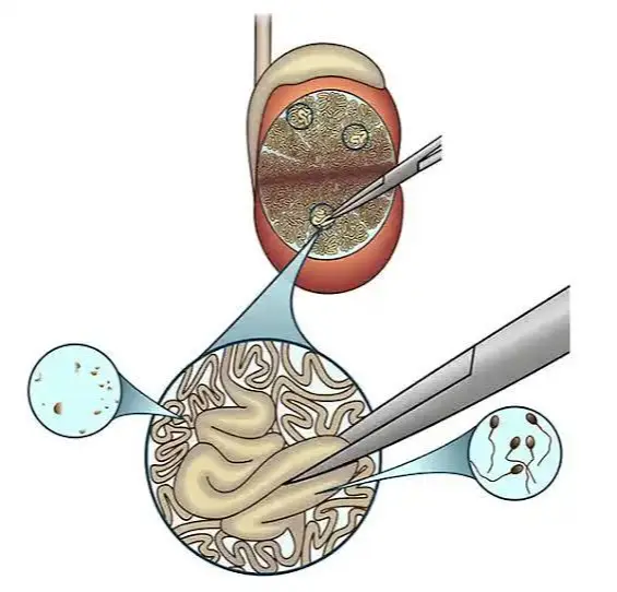
Testicular sperm retrieval (TESE):This procedure can be performed during a testicular biopsy or at another time using the same technique. After 15 years of studies, the pregnancy rate with fresh sperm obtained during biopsy was 47%, while the pregnancy rate with sperm obtained via TESE and frozen was found to be higher at 62%. TESE performed under a microscope (microTESE) has become the preferred method because it causes minimal tissue damage. During microsurgery, the surgeon can see the areas in the testicle where sperm production takes place, which is impossible with standard TESE.
Testicular sperm aspiration (TESA):Although less invasive, it yields fewer sperm compared to TESE and has no place in non-obstructive azoospermia.
Microsurgical sperm aspiration from the epididymis (MESA):This method involves directly obtaining sperm from the epididymis. Sperm within the epididymis are more mature. Under a microscope, the epididymis is opened, and a thin needle is used to aspirate the fluid from within the epididymis into a syringe containing human tubal fluid. MESA (Methodontic Sperm Aspiration) is another method of directly obtaining sperm from the epididymis. It can be performed in an outpatient setting under local anesthesia. Although effective in obtaining sperm, it cannot be performed from many areas and can cause blockages in the sperm ducts. In cases where sperm duct blockages cannot be surgically corrected, an artificial sperm sac can be surgically created to collect sperm. The collected sperm can then be aspirated multiple times with a needle under ultrasound guidance. This method is recommended and rarely used in cases of congenital vas deferens absence with normal sperm production. Methods using silicone artificial sperm sacs have been described but have not been found successful.
ELECTROEJACULATION:In patients who cannot achieve ejaculation (e.g., those with spinal cord injuries), ejaculation is achieved by delivering electrical stimulations starting from -5 volts to the seminal vesicles via a probe inserted rectally under general anesthesia. Since the resulting ejaculation will be retrograde (towards the bladder), a urethral catheter is placed in the urethra before the procedure, and buffering fluids are administered to prevent urine acidity. The ejaculated fluid is collected from the bladder using the catheter, and the presence of sperm is examined. Devices that stimulate the penis via vibration to induce ejaculation are also available and can be used at home without anesthesia.
8-ARTIFICIAL INSEMINATION (AI):This is the procedure commonly known as artificial insemination. It involves injecting sperm directly into the cervix (intracervical insemination) or into the uterus (intrauterine insemination). It is suitable for cases of low sperm concentration (no sperm detected in a postcoital test), low sperm motility, or unexplained infertility. Intrauterine insemination opens the way for natural fertilization in cases where cervical mucus prevents sperm passage. When used alone, the pregnancy rate is 4%, but when combined with ovulation induction, the pregnancy rate is 8-17%. Semen quality should be improved with semen processing methods before the procedure. The success rate of the treatment decreases with age, and the risk of spontaneous miscarriage during pregnancy is also higher than normal. If success is not achieved after 3-6 attempts at this procedure, IVF should be recommended.
9-ASSISTED REPRODUCTIVE TECHNIQUES:These methods are applied to individuals with severe oligospermia, azoospermia, unexplained infertility, and other diagnosed conditions that prevent fertility. In assisted reproductive techniques, donated or maternally obtained eggs are fertilized with the male partner's sperm or donated sperm. The resulting embryos are then placed into the female reproductive tract. The success rate is 15-20% with each procedure. With repeated applications, the success rate has reached 50%. The high cost of the procedure is the reason it is not preferred as a first-line treatment.
In vitro fertilization ( IVF ):In vitro fertilization (IVF) is the process of implanting an embryo, developed from an egg fertilized outside the uterus, into the uterus. It is suitable for couples where other techniques have been unsuccessful. Generally, at least 50,000 to 500,000 motile sperm are required for IVF. Before the procedure, the woman's pituitary gland is suppressed with GnRH, and then her ovaries are stimulated with medication. The ovaries are monitored with ultrasound, estrogen and prolactin hormone levels are measured, and when sufficient egg development is achieved, the developed eggs are retrieved vaginally. Approximately 12 eggs are collected in each ovulation cycle and immediately stored in a special container. After 3-6 hours, approximately 100,000 sperm are added to the medium for each egg. After 48 hours, embryos consisting of 3 to 8 cells develop. 2 to 4 embryos are implanted into the uterus, and the remaining embryos are frozen for later use. Pregnancy rates range from 10-45%. IVF is a safe and convenient procedure. Risks include multiple pregnancies and ovarian hyperstimulation syndrome, and the risk of major birth defects is slightly higher than normal. The incidence of hypospadias in male children born via IVF is 1.5%, compared to 0.3% in naturally born male children. This is thought to be due to the use of high doses of progesterone in the mother during egg retrieval.
Intrafallopian gamete transfer (GIFT) and intrafallopian zygote transfer (ZIFT):It is the process of directly introducing semen (GIFT) or a fertilized egg in its zygote stage (ZIFT), before it develops into an embryo, into the fallopian tubes of the female through either an open or closed surgical procedure.
The success rate of these techniques is 25-30%. However, these procedures are performed under general anesthesia and carry some risks. Fertilization and implantation in the uterus are not guaranteed, and they cannot be used in women with blocked fallopian tubes. These procedures are rarely needed as a treatment option.
Intracytoplasmic sperm injection (ICSI):This involves the direct injection of sperm into the egg under microscopic vision. It is used in cases of infertility due to severe disorders, including patients for whom simpler methods have been unsuccessful and in whom sperm has been obtained directly from the testes and epididymis.
Sperm samples are obtained through masturbation or surgically using sperm retrieval methods. In patients with high rates of necrosospermia (immobile sperm), sperm retrieval methods can also be used because sperm obtained through masturbation may contain a high percentage of damaged DNA. Sperm can be evaluated microscopically by testing their motility, shape, DNA quality, and binding capacity to hyaluronic acid. The embryologist then selects the best sperm for processing.
Oocytes (egg cells) are treated with a substance called hyaluronidase, and the surrounding cross-section is removed. A pipette is used to hold the egg in place, while sperm is transferred into the oocyte using a fine-tipped needle. Sperm is introduced at the 3 o'clock position when the egg's polar body is at the 6 or 12 o'clock position, taking care not to damage the oocyte's DNA. After 48 hours, the embryo is transferred to the uterus.
A study with 1409 oocytes found a fertilization rate of 59% and a pregnancy rate of 35%. It is known that the success rate does not differ between fresh and frozen sperm. In men undergoing ICSI due to infertility, a decrease in egg reserve in their partners reduces the success rate of TESE.
10-OTHER ASSESSMENTS
Genetic expert:If a genetic cause of infertility is known or suspected, and non-obstructive azoospermia or severe oligospermia is present, evaluation by a genetic specialist is necessary. Furthermore, in the era of ICSI and IVF, evaluation of chromosomal abnormalities that can be genetically passed on to children is crucial. Karyotype analysis (46, XY) from blood and evaluation of Y chromosome microdeletions using PCR technique should be performed. Chromosomal abnormalities are seen in 13-17% of patients with non-obstructive azoospermia. Of these, 4-16% are Klinefelter syndrome (47, XXY) and 9% are Y chromosome microdeletions.
In bilateral congenital absence of the vas deferens, a CTFR gene mutation is observed. In 50-82% of these patients, if only one gene is mutated, the genital form of cystic fibrosis occurs without respiratory problems. Patients with mutations in both genes also experience pulmonary manifestations of cystic fibrosis, although medical advances have increased their life expectancy. Therefore, assisted reproductive techniques are needed for patients with cystic fibrosis. In such patients, genetic evaluation of the female partner to determine if she is a carrier of the CTFR gene mutation is extremely important in determining whether the child will have cystic fibrosis.
Endocrinology specialist:Hormonal evaluation should be performed in patients diagnosed with severe oligospermia or azoospermia. In cases of unexplained hypogonadysfunction or elevated prolactin levels, CT or MRI should be used to check for pituitary tumors.
Diet:Antioxidants like vitamins C and E improve sperm quality by reducing the amount of free radicals that can damage cell membranes. Studies also suggest that zinc, fish oil, and selenium have beneficial effects.
Activity: Smoking, marijuana use, anabolic steroid use.Patients should be informed about the harmful effects of intercourse on sperm and advised to restrict it. They should be encouraged to avoid harmful environmental chemicals. Patients should also be told that the optimal time for intercourse to conceive is every two days, in the middle of a woman's menstrual cycle.