









Fusion Biopsy
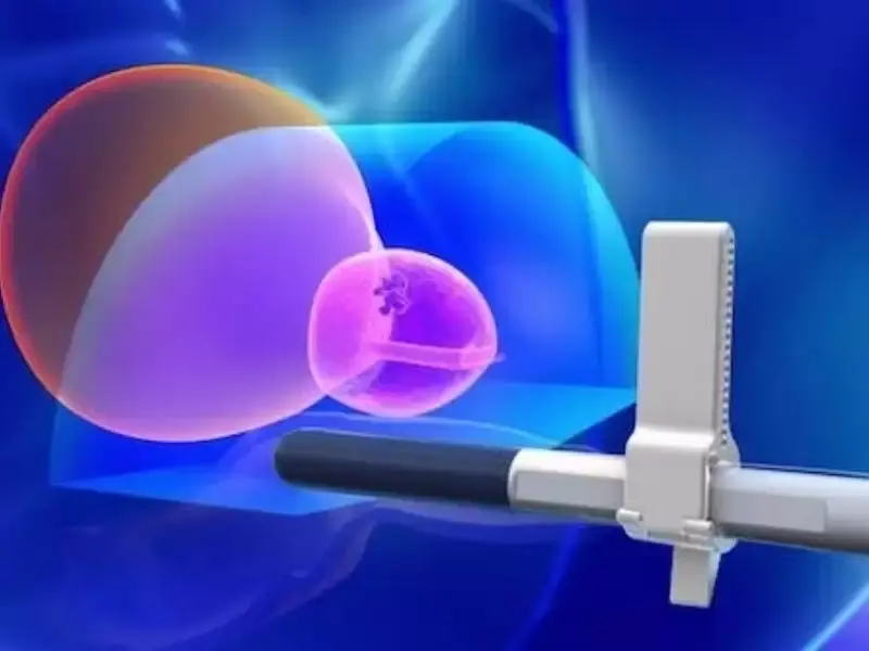
Fusion biopsy is a technique that goes beyond traditional biopsy methods and has become revolutionary especially in the diagnosis of prostate cancer. This method enables targeted biopsy by combining magnetic resonance imaging (MRI) with ultrasound images. In this way, cancerous tissues can be detected more accurately and unnecessary biopsy procedures can be avoided.
Fusion biopsy is a technique that goes beyond traditional biopsy methods and has become revolutionary especially in the diagnosis of prostate cancer. This method enables targeted biopsy by combining magnetic resonance imaging (MRI) with ultrasound images. In this way, cancerous tissues can be detected more accurately and unnecessary biopsy procedures can be avoided.
What makes this method unique is that the combination of two different imaging techniques allows the patient’s anatomy to be visualized much more clearly. MRI provides a detailed map of soft tissues, while ultrasound offers real-time guidance. When combined, these two technologies help establish a precise diagnosis, especially in patients suspected of having prostate cancer.
When biopsies are performed using conventional methods, it may not always be possible to accurately identify the target area. However, thanks to fusion biopsy, cancerous regions can be detected more precisely, reducing unnecessary tissue sampling and therefore decreasing the patient’s level of discomfort.
The Importance of Fusion Biopsy
Fusion biopsy not only increases diagnostic accuracy but also significantly improves patient comfort. Detecting cancerous tissues with this method allows the treatment process to begin earlier and increases the likelihood of successful outcomes. Early diagnosis is especially vital in diseases such as prostate cancer, which may progress silently.
In addition, fusion biopsy reduces the number of unnecessary biopsies, minimizing possible complications and infection risks to which patients are exposed. In traditional biopsy methods, doctors generally perform random tissue sampling, whereas fusion biopsy focuses on specific targets. This increases the accuracy of biopsy results.
Furthermore, fusion biopsy enables more efficient use of medical resources. Reducing unnecessary procedures eases the burden on healthcare systems and improves the overall quality of patient care. In this way, not only individual patient health but also public health is positively affected.
The Use of Fusion Biopsy in Prostate Cancer
Prostate cancer is one of the most common types of cancer in men, and early diagnosis greatly affects the success of the treatment process. At this point, fusion biopsy emerges as a revolutionary tool in the diagnosis of prostate cancer. Unlike traditional methods, this technique enables more accurate and reliable detection of cancerous tissues.
MRI fusion biopsy allows targeted biopsy in patients suspected of having prostate cancer. Through MRI scans, abnormal areas in the prostate gland are clearly identified, and samples are taken from these areas with ultrasound guidance. This significantly reduces the risk of accidentally missing cancerous tissues.
This type of biopsy is especially beneficial for high-risk patients. Patients with high PSA levels or abnormal digital rectal examination results are evaluated in greater detail with fusion biopsy. This helps physicians make more strategic treatment decisions.
MRI Fusion Biopsy: The Role of Technology
MRI fusion biopsy is one of the most advanced technologies offered by modern medicine. This method combines the detailed anatomical data of magnetic resonance imaging with the real-time imaging capability of ultrasound, providing patients with a better diagnostic opportunity. This advanced level of technology plays a critical role, especially in cancers that are difficult to detect.
MRI technology enables clear visualization of the prostate gland. Thanks to the superior resolution of this method, a detailed map of the cancerous region and surrounding tissues can be created. When this information is combined with the real-time guidance provided by ultrasound during biopsy, precise targeting can be achieved.
This technological combination not only improves diagnostic processes but also provides a significant advantage in treatment planning. The data obtained through fusion biopsy enables more effective planning of treatment options such as surgical intervention or radiotherapy. This improves patients’ quality of life and increases treatment success rates.
What Is Targeted Prostate Biopsy?
Targeted prostate biopsy is an advanced method used in the diagnosis of prostate cancer and, unlike traditional biopsy techniques, allows tissue samples to be taken from specifically identified areas. This method helps accurately identify suspicious regions and establish a correct diagnosis.
Fusion biopsy is one of the most effective applications of targeted prostate biopsy. By combining MRI and ultrasound images, doctors can better visualize abnormal regions in the prostate gland and obtain samples from these areas. This helps prevent both false-negative results and unnecessary biopsies.
Targeted biopsy is especially used in cases where previous biopsy results are inconclusive or PSA levels remain persistently high. The high accuracy provided by this method allows treatment planning to be carried out more effectively. In this way, early detection of cancerous tissues and initiation of treatment become easier.
Advantages of Fusion Biopsy
Fusion biopsy offers many advantages compared to traditional biopsy methods. First of all, this method improves patients’ quality of life. Because it is less invasive and provides accurate results, patients are protected from unnecessary procedures and complication risks.
This type of biopsy increases diagnostic accuracy. The combination of MRI and ultrasound enables doctors to detect diseases such as prostate cancer earlier and more reliably. Early diagnosis is a factor that directly affects the success of treatment and improves patients’ prognosis.
In addition, fusion biopsy is also economically advantageous. Preventing unnecessary biopsies and false-positive results significantly reduces healthcare costs. This provides long-term benefits for both individual patients and healthcare systems. The effective use of this technology is one of the most important innovations offered by modern medicine.
Risks and Limitations of Fusion Biopsy
As with every medical procedure, fusion biopsy also involves certain risks and limitations. First of all, this method may not be available in every hospital or clinic because it requires advanced technology, which means additional costs. This situation may limit access and prevent some patients from benefiting from these advantages.
During fusion biopsy, there is a risk of infection, as with conventional biopsies. However, this risk can usually be minimized through proper sterilization and preventive antibiotic treatments. Nevertheless, due to the invasive nature of the procedure, side effects such as hematoma or bleeding may occur in some patients.
Among the limitations is the inability to detect very small or deeply located tumors. MRI fusion biopsy may be less effective in certain areas of the prostate, which may require the use of traditional methods in some cases. However, these limitations can generally be overcome through technological advancements and medical expertise.
The Fusion Biopsy Process: Stages and Preparation
The fusion biopsy process requires careful planning and preparation. The first stage involves performing MRI scans of the patient. MRI scans provide a detailed image of the prostate gland, and these images are recorded for use during the procedure.
In the second stage, the biopsy procedure is performed under ultrasound guidance. By combining MRI and ultrasound images, doctors obtain tissue samples from the targeted regions. During this stage, local anesthesia is applied to ensure patient comfort. The procedure is generally short, and the patient is usually discharged on the same day.
After the procedure, the tissue samples are sent for pathological examination. The results are usually available within a few days, and the treatment plan is determined accordingly. Fusion biopsy is a critical step for accurate diagnosis and effective treatment, and every stage of this process should be carried out meticulously.
The information here is for general education only and is not medical advice. Please consult your physician for personalized treatment.